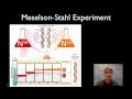
The Meselson–Stahl experiment was an experiment by Matthew Meselson and Franklin Stahl with some additional help from a Canadian biologist, Mason MacDonald, and Canadian nuclear physicist, Amandeep Sehmbi, in 1958 which supported the hypothesis that DNA replication was semiconservative. In semiconservative replication, when the double stranded DNA helix is replicated, each of the two new double-stranded DNA helices consisted of one strand from the original helix and one newly synthesized. It has been called "the most beautiful experiment in biology."[1] Meselson and Stahl decided the best way to tag the parent DNA would be to change one of the atoms in the parent DNA molecule. Since nitrogen is found in the nitrogenous bases of each nucleotide, they decided to use an isotope of nitrogen to distinguish between parent and newly copied DNA. The isotope of nitrogen had an extra neutron in the nucleus, which made it heavier.
Nitrogen is a major constituent of DNA. 14N is by far the most abundant isotope of nitrogen, but DNA with the heavier (but non-radioactive) 15N isotope is also functional.
E. coli was grown for several generations in a medium containing NH4Cl with 15N. When DNA is extracted from these cells and centrifuged on a salt density gradient, the DNA separates out at the point at which its density equals that of the salt solution. The DNA of the cells grown in 15N medium had a higher density than cells grown in normal 14N medium. After that, E. coli cells with only 15N in their DNA were transferred to a 14N medium and were allowed to divide; the progress of cell division was monitored by microscopic cell counts and by colony assay.
DNA was extracted periodically and was compared to pure 14N DNA and 15N DNA. After one replication, the DNA was found to have intermediate density. Since conservative replication would result in equal amounts of DNA of the higher and lower densities (but no DNA of an intermediate density), conservative replication was excluded. However, this result was consistent with both semiconservative and dispersive replication. Semiconservative replication would result in double-stranded DNA with one strand of 15N DNA, and one of 14N DNA, while dispersive replication would result in double-stranded DNA with both strands having mixtures of 15N and 14N DNA, either of which would have appeared as DNA of an intermediate density.
The authors continued to sample cells as replication continued. DNA from cells after two replications had been completed was found to consist of equal amounts of DNA with two different densities, one corresponding to the intermediate density of DNA of cells grown for only one division in 14N medium, the other corresponding to DNA from cells grown exclusively in 14N medium. This was inconsistent with dispersive replication, which would have resulted in a single density, lower than the intermediate density of the one-generation cells, but still higher than cells grown only in 14N DNA medium, as the original 15N DNA would have been split evenly among all DNA strands. The result was consistent with the semiconservative replication hypothesis.
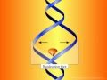
In this animation, we demonstrate the biology of DNA replication leading to bacterial cell division in a gram positive bacterium, such as S. pneumoniae. The DNA is shown as a circular double strand within the bacterial cell. Like the DNA of all living organisms, it contains the unique genetic code for all of the proteins required for bacterial survival. Bacteria replicate by a process known as binary fission whereby one bacterium separates into 2 new daughter cells. However, before this can occur, the bacterium must make an identical copy of its complete circular DNA.
In this animation, we demonstrate the biology of DNA replication leading to bacterial cell division in a gram positive bacterium, such as S. pneumoniae. The DNA is shown as a circular double strand within the bacterial cell. Like the DNA of all living organisms, it contains the unique genetic code for all of the proteins required for bacterial survival. Bacteria replicate by a process known as binary fission whereby one bacterium separates into 2 new daughter cells. However, before this can occur, the bacterium must make an identical copy of its complete circular DNA. DNA replication requires that the two strands of DNA separate so that the genetic code of the bacterium can be read and a new complimentary strand can be created for each of the original strands. To accomplish this, various enzymes known as helicases break the hydrogen bonds between the bases in the two DNA strands, unwind the strands from each other, and stabilize the exposed single strands, preventing them from joining back together. The points at which the two strands of DNA separate to allow replication of DNA are known as replication forks. The enzymes DNA polymerase then move along each strand of DNA, behind each replication fork synthesizing new DNA strands (in red) complementary to the original ones. As the replication forks move forward, positive superhelical twists in the DNA begin to accumulate ahead of them. In order for DNA replication to continue, these superhelical twists must be removed. The bacterial enzyme, DNA gyrase, which is also known as topoisomerase II, is responsible for removing the positive superhelical twists so that DNA replications can procede. DNA gyrase is an essential bacterial enzyme composed of two A and two B subunits which are products of the gyrA and gyrB genes. This enzyme has other important functions which affect the initiation of DNA replication and transcription of many genes. With the combined involvement of these enzymes, an entire duplicate copy of the bacterial genome is produced as the 2 replication forks move in opposite directions around the circular DNA genome. Eventually, as the 2 replication forks meet, 2 new complete chromosomes have been made, each consisting of 1 old and 1 new strand of DNA. This is referred to as semi-conservative replication. In order to allow the 2 new interlinked chromosomes to come apart, another bacterial enzyme is needed which is known as topoisomerase IV. This enzyme is structurally related to DNA gyrase and is coded for by the parC and parE genes. Topoisomerase IV allows for the 2 new inter-linked chromosomes to separate so that they can be segregated into 2 new daughter bacterial cells. Complete separation of bacterial cells Fluoroquinolones. First mechanism of action -- inhibition of DNA gyrase. Fluoroquinolones act by inhibiting the activity of both the DNA gyrase and the topoisomerase IV enzymes. For most gram negative bacteria, DNA gyrase is the primary fluoroquinolone target. Fluoroquinolones have been shown to bind specifically to the complex of DNA gyrase and DNA rather than to DNA gyrase alone. As a result of this binding, quinolones appear to stabilize the enzyme-DNA complexes which in turn results in breaks in the DNA that are fatal to the bacterium. A second mechanism of fluoroquinolone action is shown here. With some exceptions, topoisomerase IV is the primary target of fluoroquinolone action in most gram positive bacteria such as Staphylococci and Streptococci, with DNA gyrase being a secondary target. The separation of 2 new interlinked daughter strands of circular DNA is disrupted. The final result on the bacteria, however, is the same. Bacterial replication is disrupted and the bacterium breaks apart. The relative potency of different fluoroquinolone antibiotics (and thus their spectrum of activity) is dependent in part on their affinity for either DNA gyrase or topoisomerase IV or both. One of the most common mechanisms by which bacteria acquire resistance to fluoroquinolones is by spontaneously occurring mutations in chromosomal genes that alter the target enzymes -- DNA gyrase and topoisomerase IV or both. The frequency with which these spontaneous mutations occurs may be in the range of 10-6. The effect of mutations on the activity of an individual fluoroquinolone will vary depending on the number of mutations, the location of the mutations and which target enzyme is affected. If a mutation occurs (either in the gyrA or gyrB gene) that alters DNA gyrase and results in a reduced affinity of the fluoroquinolone antibiotic for this enzyme, the organism will become resistant.
DNA replication is the process of copying a double-stranded DNA molecule to form two double-stranded molecules. The process of DNA replication is a fundamental process used by all living organisms as it is the basis for biological inheritance. As each DNA strand holds the same genetic information, both strands can serve as templates for the reproduction of the opposite strand. The template strand is preserved in its entirety and the new strand is assembled from nucleotides — this process is called "semiconservative replication". The resulting double-stranded DNA molecules are identical; proofreading and error-checking mechanisms exist to ensure near perfect fidelity.
DNA Replication DNA Replication 2
Dna replication 3
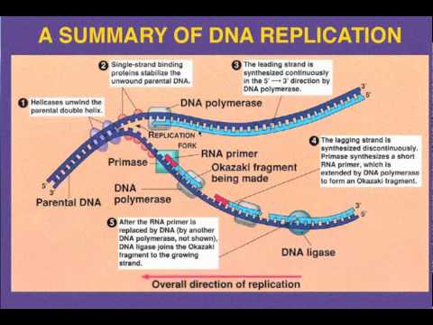
In a cell, DNA replication must happen before cell division can occur. DNA synthesis begins at specific locations in the genome, called "origins", where the two strands of DNA are separated. RNA primers attach to single stranded DNA and DNA polymerase extends from the primers to form new strands of DNA, adding nucleotides matched to the template strand. The unwinding of DNA and synthesis of new strands forms a replication fork. In addition to DNA polymerase, a number of enzymes are associated with the fork and assist in the initiation and continuation of DNA synthesis.
DNA replication can also be performed artificially, using the same enzymes used within the cell. DNA polymerases and artificial DNA primers are used to initiate DNA synthesis at known sequences in a template molecule. The polymerase chain reaction (PCR), a common laboratory technique, employs artificial synthesis in a cyclic manner to rapidly and specifically amplify a target DNA fragment from a pool of DNA.
The leading strand is the template strand of the DNA double helix so that the replication fork moves along it in the 3' to 5' direction. This allows the newly synthesized strand complementary to the original strand to be synthesized 5' to 3' in the same direction as the movement of the replication fork.
On the leading strand, a polymerase "reads" the DNA and adds nucleotides to it continuously. This polymerase is DNA polymerase III (DNA Pol III) in prokaryotes and presumably Pol ε in yeasts. In human cells the leading and lagging strands are synthesized by Pol α and Pol δ within the nucleus and Pol γ in the mitochondria. Pol ε can substitute for Pol δ in special circumstances.
Lagging strand
The lagging strand is the strand of the template DNA double helix that is oriented so that the replication fork moves along it in a 5' to 3' manner. Because of its orientation, opposite to the working orientation of DNA polymerase III, which moves on a template in a 3' to 5' manner, replication of the lagging strand is more complicated than that of the leading strand.
On the lagging strand, primase "reads" the DNA and adds RNA to it in short, separated segments. In eukaryotes, primase is intrinsic to Pol α. DNA polymerase III or Pol δ lengthens the primed segments, forming Okazaki fragments. Primer removal in eukaryotes is also performed by Pol δ.[18] In prokaryotes, DNA polymerase I "reads" the fragments, removes the RNA using its flap endonuclease domain (RNA primers are removed by 5'-3' exonuclease activity of polymerase I [weaver, 2005], and replaces the RNA nucleotides with DNA nucleotides (this is necessary because RNA and DNA use slightly different kinds of nucleotides). DNA ligase joins the fragments together.
DNA helicase is an enzyme that aids in DNA synthesis by 'unzipping' the two strands of a DNA helix so that DNA polymerase can access the DNA to add nucleotides and effect copying.
Many cellular processes (DNA replication, RNA transcription, DNA recombination, DNA repair, Ribosome biogenesis) involve the separation of nucleic acid strands. Helicases are often utilized to separate strands of a DNA double helix or a self-annealed RNA molecule using the energy from ATP or GTP hydrolysis. They move incrementally along one nucleic acid strand of the duplex with a directionality specific to each particular enzyme. There are many helicases (14 confirmed in E. coli, 24 in human cells) resulting from the great variety of processes in which strand separation must be catalyzed.
Function
Helicases adopt different structures and oligomerization states. Whereas DnaB-like helicases unwind DNA as donut shaped hexamers, other enzymes have been shown to be active as monomers or dimers. Recent studies showed that helicases do not merely wait passively for the fork to widen, but play an active role in forcing the fork to open, thus "it is an active unwinding motor". However, the unwinding is much faster in cells than in the test tube, so "accessory proteins are helping the helicase out by destabilizing the fork junction".
Structural features
The common function of helicases accounts for the fact that they display a certain degree of amino acid sequence homology; they all possess common sequence motifs located in the interior of their primary sequence. These are thought to be specifically involved in ATP binding, ATP hydrolysis and translocation on the nucleic acid substrate. The variable portion of the amino acid sequence is related to the specific features of each helicase.
Based on the presence of defined helicase motifs, it is possible to attribute a putative helicase activity to a given protein, though the presence of a motif does not confirm the protein as a helicase. Conserved motifs do, however, support an evolutionary homology among enzymes. Based on the presence and the form of helicase motifs, helicases have been separated in 4 superfamilies and 2 smaller families. Some members of these families are indicated, with the organism from which they are extracted, and their function.
Superfamilies
Superfamily I: UvrD (E. coli, DNA repair), Rep (E. coli, DNA replication), PcrA (Staphylococcus aureus, Bacillus anthracis and Bacillus cereus, regulation of recombination by displacing RecA from DNA and inhibiting RecA-mediated DNA strand exchange), Dda (bacteriophage T4, replication initiation).
Superfamily II: RecQ (E. coli, DNA repair), eIF4A (Baker's Yeast, RNA translation), WRN (human, DNA repair), NS3 (Hepatitis C virus, replication). TRCF (Mfd) (E.coli, transcription-repair coupling factor).
Mutations are changes to the base pair sequence of genetic material (either DNA or RNA). Mutations can be caused by copying errors in the genetic material during cell division and by exposure to ultraviolet or ionizing radiation, chemical mutagens, or viruses, or can occur deliberately under cellular control during processes such as meiosis or hypermutation. In multicellular organisms, mutations can be subdivided into germline mutations, which can be passed on to descendants, and somatic mutations. The somatic mutations cannot be transmitted to descendants in animals. Plants sometimes can transmit somatic mutations to their descendants asexually or sexually (in case when flower buds develop in somatically mutated part of plant).
Mutations create variation in the gene pool, and the less favorable (or deleterious) mutations are removed from the gene pool by natural selection, while more favorable (beneficial or advantageous) ones tend to accumulate, resulting in evolutionary change. For example, a butterfly may develop offspring with a new mutation caused say by ultraviolet light from the sun. In most cases, this mutation is not good, since obviously there was no 'purpose' for such change at the molecular level. However, sometimes a mutation may change, say, the butterfly's color, making it harder for predators to see it; this is an advantage and the chances of this butterfly surviving and producing its own offspring are a little better, and over time the number of butterflies with this mutation may form a large percentage of the species. Neutral mutations are defined as mutations whose effects do not influence the fitness of either the species or the individuals who make up the species. These can accumulate over time due to genetic drift. The overwhelming majority of mutations have no significant effect, since DNA repair is able to mend most changes before they become permanent mutations, and many organisms have mechanisms for eliminating otherwise permanently mutated somatic cells.
Point mutations, often caused by chemicals or malfunction of DNA replication, exchange a single nucleotide for another. Most common is the transition that exchanges a purine for a purine (A ↔ G) or a pyrimidine for a pyrimidine, (C ↔ T). A transition can be caused by nitrous acid, base mispairing, or mutagenic base analogs such as 5-bromo-2-deoxyuridine (BrdU). Less common is a transversion, which exchanges a purine for a pyrimidine or a pyrimidine for a purine (C/T ↔ A/G). A point mutation can be reversed by another point mutation, in which the nucleotide is changed back to its original state (true reversion) or by second-site reversion (a complementary mutation elsewhere that results in regained gene functionality). These changes are classified as transitions or transversions. An example of a transversion is adenine (A) being converted into a cytosine (C). There are also many other examples that can be found. Point mutations that occur within the protein coding region of a gene may be classified into three kinds, depending upon what the erroneous codon codes for: Silent mutations: which code for the same amino acid. Missense mutations: which code for a different amino acid. Nonsense mutations: which code for a stop and can truncate the protein. Insertions add one or more extra nucleotides into the DNA. They are usually caused by transposable elements, or errors during replication of repeating elements (e.g. AT repeats). Insertions in the coding region of a gene may alter splicing of the mRNA (splice site mutation), or cause a shift in the reading frame (frameshift), both of which can significantly alter the gene product. Insertions can be reverted by excision of the transposable element.
Deletions remove one or more nucleotides from the DNA. Like insertions, these mutations can alter the reading frame of the gene. They are irreversible.
Causes of mutation
Two classes of mutations are spontaneous mutations (molecular decay) and induced mutations caused by mutagens.
Spontaneous mutations on the molecular level include: Tautomerism - A base is changed by the repositioning of a hydrogen atom. Depurination - Loss of a purine base (A or G). Deamination - Changes a normal base to an atypical base; C → U, (which can be corrected by DNA repair mechanisms), or spontaneous deamination of 5-methycytosine (irreparable), or A → HX (hypoxanthine). Transition - A purine changes to another purine, or a pyrimidine to a pyrimidine. Transversion - A purine becomes a pyrimidine, or vice versa.
Benzopyrene, the major mutagen in tobacco smoke, in an adduct to DNA. Produced from PDB 1JDG.
Induced mutations on the molecular level can be caused by:
Chemicals
Nitrosoguanidine (NTG)
Hydroxyamine NH3OH
Base analogs (e.g. BrdU)
Simple chemicals (e.g. acids)
Alkylating agents (e.g. N-ethyl-N-nitrosourea (ENU)) These agents can mutate both replicating and non-replicating DNA. In contrast, a base analog can only mutate the DNA when the analog is incorporated in replicating the DNA. Each of these classes of chemical mutagens has certain effects that then lead to transitions, transversions, or deletions.
Methylating agents (e.g. ethyl methanesulfonate (EMS))
Polycyclic hydrocarbons (e.g. benzopyrenes found in internal combustion engine exhaust)
DNA intercalating agents (e.g. ethidium bromide)
DNA crosslinker (e.g. platinum)
Oxidative damage caused by oxygen(O)] radicals
Radiation
Ultraviolet radiation (nonionizing radiation) - excites electrons to a higher energy level. DNA absorbs one form, ultraviolet light. Two nucleotide bases in DNA - cytosine and thymine-are most vulnerable to excitation that can change base-pairing properties. UV light can induce adjacent thymine bases in a DNA strand to pair with each other, as a bulky dimer.
Ionizing radiation
DNA has so-called hotspots, where mutations occur up to 100 times more frequently than the normal mutation rate. A hotspot can be at an unusual base, e.g., 5-methylcytosine.
Mutation rates also vary across species. Evolutionary biologists have theorized that higher mutation rates are beneficial in some situations, because they allow organisms to evolve and therefore adapt more quickly to their environments. For example, repeated exposure of bacteria to antibiotics, and selection of resistant mutants, can result in the selection of bacteria that have a much higher mutation rate than the original population (mutator strains).
Harmful mutations
Changes in DNA caused by mutation can cause errors in protein sequence, creating partially or completely non-functional proteins. To function correctly, each cell depends on thousands of proteins to function in the right places at the right times. When a mutation alters a protein that plays a critical role in the body, a medical condition can result. A condition caused by mutations in one or more genes is called a genetic disorder. However, only a small percentage of mutations cause genetic disorders; most have no impact on health. For example, some mutations alter a gene's DNA base sequence but don’t change the function of the protein made by the gene.
If a mutation is present in a germ cell, it can give rise to offspring that carries the mutation in all of its cells. This is the case in hereditary diseases. On the other hand, a mutation can occur in a somatic cell of an organism. Such mutations will be present in all descendants of this cell, and certain mutations can cause the cell to become malignant, and thus cause cancer.
Often, gene mutations that could cause a genetic disorder are repaired by the DNA repair system of the cell. Each cell has a number of pathways through which enzymes recognize and repair mistakes in DNA. Because DNA can be damaged or mutated in many ways, the process of DNA repair is an important way in which the body protects itself from disease.
Beneficial mutations
A very small percentage of all mutations actually have a positive effect. These mutations lead to new versions of proteins that help an organism and its future generations better adapt to changes in their environment. For example, a specific 32 base pair deletion in human CCR5 (CCR5-32) confers HIV resistance to homozygotes and delays AIDS onset in heterozygotes.[1] The CCR5 mutation is more common in those of European descent. One theory for the etiology of the relatively high frequency of CCR5-32 in the European population is that it conferred resistance to the bubonic plague in mid-14th century Europe.