Ehlers-Danlos syndrome is a group of disorders that affect connective tissues, which are tissues that support the skin, bones, blood vessels, and other organs. Defects in connective tissues cause the signs and symptoms of Ehlers-Danlos syndrome, which vary from mildly loose joints to life-threatening complications.
In 1997, researchers proposed a simpler classification that reduced the number of major types to six and gave them descriptive names: the arthrochalasia type, the classic type, the dermatosparaxis type, the hypermobility type, the kyphoscoliosis type, and the vascular type. Other forms of the condition may exist, but they have been reported only in single families or are not well characterized.
Although all types of Ehlers-Danlos syndrome affect the joints and many also affect the skin, features vary by type. An unusually large range of joint movement (hypermobility) occurs with most forms of Ehlers-Danlos syndrome, particularly the hypermobility type. Infants with hypermobile joints often appear to have weak muscle tone, which can delay the development of motor skills such as sitting, standing, and walking. The loose joints are unstable and prone to dislocation, chronic pain, and early-onset arthritis. Dislocations involving both hips are a characteristic finding in infants with the arthrochalasia type of Ehlers-Danlos syndrome.
Causes
Mutaions in ADAMTS2, COL1A1, COL1A2, COL3A1, COL5A1, COL5A2, PLOD1, and TNXB genes cause Ehlers-Danlos syndrome.Researchers have identified more than 20 mutations in PLOD1 gene in affected persons,These mutations cause a form of Ehlers-Danlos syndrome called the kyphoscoliosis type,The most common mutation duplicates a large portion of the gene, resulting in the production of a nonfunctional version of the lysyl hydroxylase 1 enzyme. Several other mutations introduce premature stop signals that prevent the gene from making any functional enzyme. A loss of lysyl hydroxylase 1 activity impairs cross-linking between collagen molecules. This disruption in the network of collagen fibrils weakens connective tissues, causing the signs and symptoms of Ehlers-Danlos syndrome.
Breast cancer is one of common malignancy in women and second leading cause of cancer death in women after lung cancer, anatomically the breast is consist of nipple, areota, lactiferous sinus, lactiferous ducts and lactating lobular unit, breast cancer can be caused by hormonal or genetic factors, there are several types of breast cancer depending on site of origin and microscopic features, ex lobular carcinoma, invasive carcinoma, ductal carcinoma invasive ductal carcinoma and papillary carcinoma,
Lobular carcinoma starts in parts of the breast, called lobules that produce milk.In rare cases, breast cancer can start in other areas of the breast. Ductal carcinoma starts in the tubes (ducts) that move milk from the breast to the nipple. Most breast cancers are of this type.
Genes -- Some people have genes that make them more prone to developing breast cancer. The most common gene defects are found in the BRCA1 and BRCA2 genes. These genes normally produce proteins that protect you from cancer. But if a parent passes you a defective gene, you have an increased risk for breast cancer. Women with one of these defects have up to an 80% chance of getting breast cancer sometime during their life.
Symptoms
Breast lump or lump in the armpit that is hard, has uneven edges, and usually does not hurt Change in the size, shape, or feel of the breast or nipple -- for example, you may have redness, dimpling, or puckering that looks like the skin of an orange Fluid coming from the nipple -- may be bloody, clear-to-yellow, or green, and look like pus Men get breast cancer, too. Symptoms include breast lump and breast pain and tenderness.
Breast cancer can spread locally through the lymphatic system to draining lymph nodes nearby and distally through blood to the brain, liver, bone, and lung
Treatment
Breast cancer can be diagnosed by self-examination of the breast. Mammography and biopsy, Breast cancer can be treated by surgery, hormonal therapy and chemotherapy or some combination of these treatments.
At present treatment for HIV infection involves using a combination of drugs. One type of drug inhibits nucleic acid replication, for example, when AZT is incorporated into DNA by reverse trasncriptase, no additional nucleotides can be added.
A second class of drugs inhibits a protease involved in cleaving large polyproteins into functional segments. When the protease is inhibited, virus particles cannot be produced Use of a combination of drugs directed at different targets reduces the likelihood of the development of resistance by mutation.
Research is underway to develop new treatments for HIV. Chemicals called chemokines appear to inhibit HIV infection by binding to and blocking the CXCR4 and CCR5 co receptors on the host cells.
A search is in progress to apply such chemicals in HIV treatment,Some individuals who are HIV positive, but did not develop AIDS have a mutation in the CCR5 receptor that makes it defective.
Research is in progress to determine whether disabling the CCR5 receptor might beeffective in controlling AIDS in infected patients.An antiviral factor called CAF is found in CD8 cells.Research is in progress to determine if this substance could be used to block replication of HIV.
There is currently no approved vaccine against HIV infection. One problem is that HIVsurface proteins such as gp120 mutate at a high rate.Scientist may be able to construct a vaccine using an HIV with a defective form of the viral gene nef.
Viruses with the defective nef gene seem to have reduced reproductive capability, allowing the immune system to keep the virus in check.
Glucocorticoid hormone predominately affects the metabolism of carbohydrates and to a lesser extent, fats and proteins, Glucocorticoid are made in the outside portion of the adrenal glands and it’s chemically classed as steroids.
Action of Glucocortocoid hormone
Glucocorticoid hormones are steroid hormones that influence nutrient metabolism, These hormones activate genes for proteins involved in process such as the synthesis of glucose, the breakdown of proteins, and the mobilization of fats.
Glucocorticoid hormones are secreted by endocrine glands into the blood stream. They enter cells through transporters in the plasma membrane, once inside the cytosol the hormone molecules can bind to glucocortocid receptors, which function as transcription factors that activate genes. The binding of the hormone causes proteins that were bound to the receptor such as the heat shock protein hsp90 to be released.
Release of HSP90allows two receptors to bind and form a dimer as well as exposing a nuclear localization signal on the receptor.The receptor dimer is then allowed to enter into the nucleus through a nuclear pore.
The receptor dimer is able to bind to a specific DNA sequence regions called glucocortocid response elements, or GREs,that are near target genes.The GREs function as enhancers, so that binding of a receptor dimer to a GRE activates transcription of the nearby target gene.
Skin break and intradermal test are two allergy testing methods ,A clinican uses a fine needle to pick a drop of allergin like pre pollen dust animal dander under top most skin layer in the skin prick test clinican injects the solution containing alergen into the skin in the interdermal test ,the redness results at the test site in allergin sensitive person is refferd to as flare ,where the swolling results from raised bump is refered to as wheal
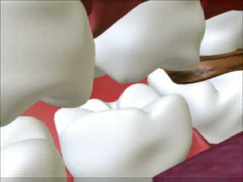
There are four main criteria required for caries formation: a tooth surface (enamel or dentin); caries-causing bacteria; fermentable carbohydrates (such as sucrose); and time.The caries process does not have an inevitable outcome, and different individuals will be susceptible to different degrees depending on the shape of their teeth, oral hygiene habits, and the buffering capacity of their saliva. Dental caries can occur on any surface of a tooth which is exposed to the oral cavity, but not the structures which are retained within the bone.
There are certain diseases and disorders affecting teeth which may leave an individual at a greater risk for caries. Amelogenesis imperfecta, which occurs between 1 in 718 and 1 in 14,000 individuals, is a disease in which the enamel does not fully form or forms in insufficient amounts and can fall off a tooth. In both cases, teeth may be left more vulnerable to decay because the enamel is not able to protect the tooth.
In most people, disorders or diseases affecting teeth are not the primary cause of dental caries. Ninety-six percent of tooth enamel is composed of minerals.These minerals, especially hydroxyapatite, will become soluble when exposed to acidic environments. Enamel begins to demineralize at a pH of 5.5. Dentin and cementum are more susceptible to caries than enamel because they have lower mineral content. Thus, when root surfaces of teeth are exposed from gingival recession or periodontal disease, caries can develop more readily. Even in a healthy oral environment, however, the tooth is susceptible to dental caries.
The anatomy of teeth may affect the likelihood of caries formation. Where the deep grooves of teeth are more numerous and exaggerated, pit and fissure caries are more likely to develop. Also, caries are more likely to develop when food is trapped between teeth.
Bacteria
The mouth contains a wide variety of bacteria, but only a few specific species of bacteria are believed to cause dental caries: Streptococcus mutans and Lactobacilli among them. Lactobacillus acidophilus, Actinomyces viscosus, Nocardia spp., and Streptococcus mutans are most closely associated with caries, partiuclarly root caries. Bacteria collect around the teeth and gums in a sticky, creamy-coloured mass called plaque, which serves as a biofilm. Some sites collect plaque more commonly than others. The grooves on the biting surfaces of molar and premolar teeth provide microscopic retention, as does the point of contact between teeth. Plaque may also collect along the gingiva. In addition, the edges of fillings or crowns can provide protection for bacteria, as can intraoral appliances such as orthodontic braces or removable partial dentures.
Fermentable carbohydrates
Bacteria in a person's mouth convert glucose, fructose, and most commonly sucrose (table sugar) into acids such as lactic acid through a glycolytic process called fermentation.If left in contact with the tooth, these acids may cause demineralization, which is the dissolution of its mineral content. The process is dynamic, however, as remineralization can also occur if the acid is neutralized by saliva or mouthwash. Fluoride toothpaste or dental varnish may aid remineralization.a If demineralization continues over time, enough mineral content may be lost so that the soft organic material left behind disintegrates, forming a cavity or hole.
Cornea is a transparent front part of the eyes that covers the iris,pupil and anterior chamber providing most of an eyes optical power
Structure of the Cornea
Although the cornea is clear and seems to lack substance, it is actually a highly organized group of cells and proteins. Unlike most tissues in the body, the cornea contains no blood vessels to nourish or protect it against infection. Instead, the cornea receives its nourishment from the tears and aqueous humor that fills the chamber behind it. The cornea must remain transparent to refract light properly, and the presence of even the tiniest blood vessels can interfere with this process. To see well, all layers of the cornea must be free of any cloudy or opaque areas.
The corneal tissue is arranged in five basic layers, each having an important function. These five layers are:
Epithelium
The epithelium is the cornea's outermost region, comprising about 10 percent of the tissue's thickness. The epithelium functions primarily to: (1) Block the passage of foreign material, such as dust, water, and bacteria, into the eye and other layers of the cornea; and (2) Provide a smooth surface that absorbs oxygen and cell nutrients from tears, then distributes these nutrients to the rest of the cornea. The epithelium is filled with thousands of tiny nerve endings that make the cornea extremely sensitive to pain when rubbed or scratched. The part of the epithelium that serves as the foundation on which the epithelial cells anchor and organize themselves is called the basement membrane.
Bowman's Layer
Lying directly below the basement membrane of the epithelium is a transparent sheet of tissue known as Bowman's layer. It is composed of strong layered protein fibers called collagen. Once injured, Bowman's layer can form a scar as it heals. If these scars are large and centrally located, some vision loss can occur.
Stroma
Beneath Bowman's layer is the stroma, which comprises about 90 percent of the cornea's thickness. It consists primarily of water (78 percent) and collagen (16 percent), and does not contain any blood vessels. Collagen gives the cornea its strength, elasticity, and form. The collagen's unique shape, arrangement, and spacing are essential in producing the cornea's light-conducting transparency.
Descemet's Membrane
Under the stroma is Descemet's membrane, a thin but strong sheet of tissue that serves as a protective barrier against infection and injuries. Descemet's membrane is composed of collagen fibers (different from those of the stroma) and is made by the endothelial cells that lie below it. Descemet's membrane is regenerated readily after injury.
The endothelium is the extremely thin, innermost layer of the cornea. Endothelial cells are essential in keeping the cornea clear. Normally, fluid leaks slowly from inside the eye into the middle corneal layer (stroma). The endothelium's primary task is to pump this excess fluid out of the stroma. Without this pumping action, the stroma would swell with water, become hazy, and ultimately opaque. In a healthy eye, a perfect balance is maintained between the fluid moving into the cornea and fluid being pumped out of the cornea. Once endothelium cells are destroyed by disease or trauma, they are lost forever. If too many endothelial cells are destroyed, corneal edema and blindness ensue, with corneal transplantation the only available therapy.
Homocystinuria, also known as Cystathionine beta synthase deficiency which occurs 1 in 200000 to 335000 people world wide,It is an inherited disorder of the metabolism of the amino acid methionine, often involving cystathionine beta synthase.This form of homocystinuria is characterized by dislocation of the lens in the eye, an increased risk of abnormal blood clots, and skeletal abnormalities. It is an inherited autosomal recessive trait, which means a child needs to inherit the defective gene from both parents to be affected.
The CBS gene provides instructions for producing cystathionine beta-synthase. This enzyme is responsible for one step in a chemical pathway that processes the amino acid methionine. Other amino acids, including homocysteine, are also products of this pathway. Mutations in the CBS gene disrupt the function of cystathionine beta-synthase, preventing homocysteine from being used properly. As a result, this amino acid and other potentially toxic substances build up in the blood, and some of the excess homocysteine is excreted in urine.
Sexual Evolution from X to Y chromosme is delivered by Dr.David C. Page M.D..He completed B.A., chemistry, Swarthmore College M.D., Harvard Medical School and Harvard–MIT Health Sciences and Technology
David Page has spent his career examining this compact chromosome: tracing its evolution, determining its nucleotide sequence, and unraveling the mechanism that allows the Y chromosome to fix its genes. His work has led to an appreciation of the complexity of the Y chromosome and to a better understanding of male infertility
Haemochromatosis is a hereditary disease characterized by excessive absorption of dietary iron resulting in a pathological increase in total body iron stores.Excess iron accumulates in tissues and organs disrupting their normal function.The hereditary form of the disease is most common among those of Northern European ancestry, in particular those of British or Irish descent.
Haemochromatosis is protean in its manifestations, i.e., often presenting with signs or symptoms suggestive of other diagnoses that affect specific organ systems. Many of the signs and symptoms below are uncommon and for most patients with the hereditary form of haemochromatosis do not show any overt signs of disease nor do they suffer premature morbidity. The more common clinical manifestations include:
* Malaise
* Liver cirrhosis (with an increased risk of hepatocellular carcinoma Liver disease is always preceded by evidence of liver dysfunction including elevated serum enzymes specific to the liver.
* Insulin resistance (often patients have already been diagnosed with diabetes mellitus type 2) due to pancreatic damage from iron deposition
* Erectile dysfunction and hypogonadism
* Decreased libido secondary to the above
* Congestive heart failure, arrhythmias or pericarditis
* Arthritis of the hands (especially the first and second MCP joints), but also the knee and shoulder joints
o Adrenal gland (leading to adrenal insufficiency)
Less common findings including:
* Deafness
* Dyskinesias, including Parkinsonian symptoms
* Dysfunction of certain endocrine organs:
o Parathyroid gland (leading to hypocalcaemia)
o Pituitary gland
* A darkish colour to the skin (see pigmentation, hence its name Diabetes bronze when it was first described by Armand Trousseau in 1865)
* An increased susceptibility to certain infectious diseases caused by siderophilic microorganisms:
o Vibrio vulnificus infections from eating seafood
o Listeria monocytogenes
o Yersinia enterocolica
o Salmonella enterica (serotype Typhymurium)
o Klebsiella pneumoniae
o Escherichia coli
o Rhizopus arrhizus
o Mucor species
Males are usually diagnosed after their forties and fifties, and women several decades later, owing to regular iron loss through menstruation (which ceases in menopause). The severity of clinical disease in the hereditary form varies considerably. There is evidence suggesting that hereditary haemochromatosis patients affected with other liver ailments such as hepatitis or alcoholic liver disease suffer worse liver disease than those with either condition alone. There are also juvenile forms of hereditary haemochromatosis that present in childhood with the same consequences of iron overload.
Genetics:
The regulation of dietary iron absorption is complex and our understanding is incomplete. One of the better characterized genes responsible for hereditary haemochromatosis is HFE on chromosome 6 which codes for a protein that participates in the regulation of iron absorption. The HFE gene has two common alleles, C282Y and H63D. Heterozygotes for either allele do not manifest clinical iron overload but may display an increased iron uptake. Mutations of the HFE gene account for 90% of the cases of non-transfusional iron overload. This gene is closely linked to the HLA-A3 locus. Homozygosity for the C282Y mutation is the most common genotype responsible for clinical iron accumulation, though heterozygosity for C282Y/H63D mutations, so-called compound heterozygotes, results in clinically evident iron overload. There is considerable debate regarding the penetrance -- the probability of clinical expression of the trait given the genotype -- is for clinical disease in HHC homozygotes. Most, if not all, males homozygous for HFE C282Y will show manifestations of liver dysfunction such as elevated liver-specific enzymes such as serum gamma glutamyltransferase (GGT) by late middle age. Homozygous females can delay the onset of iron accumulation because of iron loss through menstruation. Each patient with the susceptible genotype accumulates iron at different rates depending on iron intake, the exact nature of the mutation and the presence of other insults to the liver such as alcohol and viral disease. As such the degree to which the liver and other organs is affected, expressivity, is highly variable and is dependent on such these other factors and co-morbidities as well as age at which they are studied for manifestations of disease. Penetrance differs between different populations.
Ventilation is the first stage of Respiratory cycle followed by external respiration, internal transport and internal respiration, Ventilation has two components inspiration and expiration. The transportation of air from in and out of the lungs. The air movement is driven by changes in the pressure within the thoracic cavity, during inspiration pressure inside the cavity decreases below that of the outside air causing air to follow in to the lungs and expand the size of thoracic cage.
Sanger and co-workers developed a method for enzymatic sequencing using chain terminators it is called has Sanger sequencing or Dideoxy sequencing reactions.Fed Sanger’s method of DNA sequencing was based on Arthur Kornberg’s earlier work on DNA replication. Remember that a new DNA strand is synthesized using an existing strand as a template.
To illustrate the technique, let’s use poly-thymine DNA as an example.
The 5` carbon of an “incoming ‘ deoxynucleotide (dNTP) is joined to the 3` carbon at the end of the chain. Hydroxyl groups in each position form ester linkages with a central phosphate. In this way, the nucleotide chain elongates. The key to sangers sequencing method is the peculiar chemistry of dideoxynucleotides (didNTP). Like a deoxynucleotide, a didNTP is incorporated into a chain by forming a phosphodiester linkage at its 5` end.
Subscribe in a reader However, the didNTP lacks a 3` hydroxyl goup (OH) necessary to form the linkage with an incoming nucleotide. So the addition of a didNTP halts elongation. To sequence DNA,Four separate reactions are necessary one to provide sequence information about each of the nucleotides. Each reaction contains: template DNA, a short primer (about 20 nucleotides), DNA polymerase, and the four dNTPs (one radioactively labeled). One type of didNTP A, T, C, or G is added to each. First the DNA is denatured into single strands at nearing-boiling temperature. At high temperatures, the kinetic motion of the DNA molecule disrupts the weak hydrogen bonds that join complementary DNA strands together. When the temperature is lowered, the primer binds to its complementary sequence in the template DNA. Kornberg’s earlier work showed that a primer is needed for DNA polymerase to start replication. The DNA polymerase makes no distinction between dNTPs or didNTPs each time a didNTP is incorporated, In this case didATP, synthesis is “terminated” and a DNA strand of a discrete is generated. In this sequencing example, the didATP(purple) has terminated the reaction. The dATP happens to be the radioactive tracer but this has no effect on elongation. Because billions of DNA molecules are present, the elongation reaction can be terminated at any adenine, position. This results in collections of DNA strands of different lengths.Same thing happens to other three(G ,C,T) terminator reaction. Each reaction is then loaded into a separate lane of a polyacrylamide gel containing urea, which prevents the DNA strands from renaturing during electrophoresis. Ionized phosphates give the DNA molecule a negative charge, so DNA migrates toward the positive pole of an electric field. The movement of DNA molecules through the polyacrylamide matrix is size dependent. A blue dye that also migrates to the + pole is added to the samples to track the progress of DNA in the sequencing gel. The dye run just a little faster than the smaller fragments of DNA. Over the course of electrophoresis, shorter DNA molecules will move further down the gel than larger ones. Millions of terminated molecules of the same size will migrate to the same place and “band” in the gel. After electrophoresis the gel is sandwiched against X-ray film. The radioactive adenine in the synthesized DNA emits beta particles that expose to film, making a record of the positions of DNA bands in the gel. The sequencing gel is then read from bottom to top. The sequence of bands in the various terminator lanes gives the sequence of nucleotides in the template DNA.A typical sequencing reaction will yields 200-500 base pairs of readable sequence.
Glossary: Deoxynucleotide (dNTP) are components of DNA, containing the phosphate, sugar and organic base; when in the triphosphate form, they are the precursors required by DNA polymerase for DNA synthesis (i.e., ATP, CTP, GTP, TTP). Dideoxynucleotides (didNTP) are chain-terminating precursors of DNA synthesis that block further polymerization when added to the end of the DNA strand by DNA polymerase. These nucleotides lack a 3'-OH (hydroxyl) group necessary for continued 5'-to-3' DNA synthesis. DNA Polymerase:An enzyme that assembles new DNA by copying an existing strand. Primer: A short DNA sequence used to initiate the synthesis of DNA, as in a polymerase chain reaction. A Polyacrylamide Gel is a separation matrix used in electrophoresis of biomolecules, such as proteins or DNA fragments. Gel Electrophoresis: An Electrochemical Process in Which Charged Molecules Migrate in a gel under the Influence of an electric current.
Green florescent protein (GFP) has revolutionized research in medicine and biology, enabling scientist to get a visual fix on how organs function on the spread of disease and the response of infected cells to treatment,
In 2008 Nobel Chemsitry prize was announced to Osamu Shimomura of Japan and American duo Martin chalfie and Roger Tsien for deriving Florescent protein from a jellyfish Aequorea Victoria.
The GFP is composed of 238 amino acids; it is isolated from jellyfish Aequorea Victoria that fluoresces green when exposed to blue light.It was discovered
GFP has a typical beta barrel structure, consisting of one β-sheet with a alpha helix containing the fluorophore running through the center, while the tightly packed barrel shell protects the flurophore from quenching by the surrounding microenviornment,the inward facing side chains of the barrel induce specific cyclization reactions on the tripeptide Ser65-Tyr66-Gly67 that lead to fluorophore formation. This occurs in a series of discrete steps with distinct excitation and emission properties.
GFP has functioned has a guiding star for bichemist, biologist and medical scientist, The gene to make GFP is inserted into the DNA of lab animals, bacteria or other cells, where it is switched on by other genes, the glow becomes apparent under ultraviolet light, The telltale protein gives researchers an instant way of monitoring process that were previously invisible.
By targeting nerve cells in Alzheimer’s disease person scientist can follow the, destruction caused by disease, Tumour progression can be followed by adding GFP to cancer cells, By adding GFP to a growing mouse embryo, they can see how the pancreas generates insulin-producing beta cells. Today GFP is a standard tool for thousands of researchers all over the world
GFP derivatives
Due to the potential for widespread usage and the evolving needs of researchers, many different mutants of GFP have been engineered. The first major improvement was a single point mutation (S65T) reported in 1995 in Nature by Roger Tsien This mutation dramatically improved the spectral characteristics of GFP, resulting in increased fluorescence, photostability and a shift of the major excitation peak to 488nm with the peak emission kept at 509 nm. This matched the spectral characteristics of commonly available FITC filter sets, increasing the practicality of use by the general researcher. The addition of the 37 °C folding efficiency (F64L) point mutant to this scaffold yielded enhanced GFP (EGFP). EGFP has an extinction coefficient (denoted ε), also known as its optical cross section of 9.13×10⁻²¹ m²/molecule, also quoted as 55,000 M−1cm−1. The quantum yield (QY) of EGFP is 0.60. The relative brightness, expressed as ε•QY, is 33,000 M−1cm−1. Superfolder GFP, a series of mutations that allow GFP to rapidly fold and mature even when fused to poorly folding peptides, was reported in 2006.
Many other mutations have been made, including color mutants; in particular blue fluorescent protein(EBFP, EBFP2, Azurite, mKalama1), cyan fluorescent protein (ECFP, Cerulean, CyPet) and yellow fluorescent protein derivatives (YFP, Citrine, Venus, YPet). BFP derivatives (except mKalama1) contain the Y66H substitution. The critical mutation in cyan derivatives is the Y66W substitution, which causes the chromophore to form with an indole rather than phenol component. Several additional compensatory mutations in the surrounding barrel are required to restore brightness to this modified chromophore due to the increased bulk of the indole group. The red-shifted wavelength of the YFP derivatives is accomplished by the T203Y mutation and is due to π-electron stacking interactions between the substituted tyrosine residue and the chromophore.These two classes of spectral variants are often employed for fluorescence resonance energy transfer (FRET) experiments. Genetically-encoded FRET reporters sensitive to cell signaling molecules, such as calcium or glutamate, protein phosphorylation state, protein complementation, receptor dimerization and other processes provide highly specific optical readouts of cell activity in real time.
Semirational mutagenesis of a number of residues led to pH-sensitve mutants known as pHluorins, and later super-ecliptic pHluorins. By exploiting the rapid change in pH upon synaptic vesicle fusion, pHluorins tagged to synaptobrevin have been used to visualize synaptic activity in neurons.
The nomenclature of modified GFPs is often confusing due to overlapping mapping of several GFP versions onto a single name. For example,mGFP often refers to a GFP with an N-terminal palmitoylation that causes the GFP to bind to cell membranes. However, the same term is also used to refer to monomeric GFP, which is often achieved by the dimer interface breaking A206K mutation. Wild-type GFP has a weakdimerization tendency at concentrations above 5 mg/mL. mGFP also stands for "modified GFP" which has been optimized through amino acid exchange for stable expression in plant cells.
Use
The availability of GFP and its derivatives has thoroughly redefined fluorescence microscopy and the way it is used in cell biology and other biological disciplines. While most small fluorescent molecules such as FITC (fluorescein isothiocyanate) are strongly phototoxic when used in live cells, fluorescent proteins such as GFP are usually much less harmful when illuminated in living cells. This has triggered the development of highly automated live cell fluorescence microscopy systems which can be used to observe cells over time expressing one or more proteins tagged with fluorescent proteins. Analysis of such time lapse movies has redefined the understanding of many biological processes including protein folding, protein transport, and RNA dynamics, which in the past had been studied using fixed (i.e. dead) material.
Another powerful use of GFP is to express the protein in small sets of specific cells. This allows researchers to optically detect specific types of cells in vitro (in a dish), or even in vivo (in the living organism).
Toxins trigger the inflammatory response in the lungs,T-lymphocytes immune cells release a protein called chemokines which signal other white blood cells called neutrophylls to migrate to the lungs connective tissues and airway lumen,accumalated neutrophylls attract alveoli macrophages another white blood cell type ,both white blood cells engulf and destroy harmful particles in process called phagocytosis
The Auditory portion of inner ear is called Cochlea,organ of corti is the core component of cochlea which is the sensory organ of hearing, The structure of Cochlea consist of Scala vestibuli (containing perilymp),Scala tympani(containing perilymph),Scala media(containing endolymph),Helicotrema,Reissner's membrane,Basilar membrane ,organ of corti and hair cells,
Scala vestibulin lies superior to the cochlear duct
Scala tympani lies inferior to the scala media and terminates at the round window
Scala media is a membranous duct containing the organ of corti
Helicotrema is the location where scala tympani and scala vestiduli merge
Reissner's membrane separates the scala vestibuli from scala media
Basilar membrane separates the scala media from the scala tympani and determines the mechanical wave propagation properties of the cochlear partion
Organ of coti is the cellular layer on the basilar membrane,powered by te potential differnce the perilymph and the endolymph
Hair cells are the sensory cells in the organ of corti
>
Function
When the cochlea uncoils it will be continuous tube divided into 3 separate compartments the Scala media ,Scala tympani ,Scala vestibulin,sound waves are transmitted in scala vestibulin and reach the organ of corti on the Basilar membrane, where stimulate hair like receptor whose tips are fixed in Tectorial membrane
Light Dependent Reaction is the initial stage of Photosynthesis Where solor energy is converted into potential energy ,the light Dependent Reaction produces oxygen gas and converts ADP and NADP+ into ATP and NADPH
Process Light Dependent Reaction
Photosytem consist of rays of chlorophyll molecules, chlorophyll the green pigments of leafs absorbs light energy, and the absorbed energy excites the electrons to higher energy level. Energized electrons from photosystem 1 are passed on to electron transport chain and added to NADP+ to form NADPH, Energized electrons of photsystem 2 are passed to another electron transport chain, their energy used to pump hydrogen ions from stoma into Thylakoid compartment creating concentration gradient.
Electron leaving this electron transport chain enter photsystem 1 replenishing its lost electrons. Photosystem 2 replenished its electron by splitting water, hydrogen ions and oxygen are released in Thylakoid compartment this is were the oxygen gas generated by photosynthesis comes from, The buildup of hydrogen ions inside the thylakoid compartment stores potential energy, this energy is harvested by a enzyme called ATP synthase, as hydrogen ions diffuse through ATP synthase down the concentration gradient .the enzyme uses the energy of moving ions to make ATP, next ATP and NADPH are used in sugar making process of the Calvin cycle
Gas exchange or ventilation is one of most important process in respiration, It takes place in respiratory surface, Gas exchange is carried out by mechanism of heart and lungs,Respiration can be classified into five types namely, Breathing, External respiration, Gas Transport, Internal respiration and cellular respiration.
During external respiration oxygen and carbon dioxide are transferred between the air in lungs and the blood, alveoli that are the site of gas exchange have the walls that are 2 Cells thick, which facilitate the transport of gases by diffusion
Diffusion
Oxygen, carbon dioxide and hydrogen is carried by blood between tissues and the lungs, 70 % of CO2 is dissolved in Plasma (it is dissolved by bicarbonate;60%).Remaining 30% is transported in red blood cells combined with globin portion of Hemoglobin as carbaminohaemoglobin.
In diffusion 93% Co2 goes into red blood cells and remaining 7% is dissolved in Plasma.
Pat Brown developed a technique where cDNAs can be embedded onto lass slided.Using these DNA arrays,he could do large-scale expression studies
div style="text-align: justify;">Growth stage-specific mRNA are isoloated,and then reverse transcribed to give inique cDNA populations.These are directly embedded onto specially cated gass slides.
These slides are coated with poly-lysine,which is positively charged.DNA is negatively charged,so that cDNA "sticks" to the slide through an ionic interaction.The cDNA can still interact with DNA probe.
<
Brown used srrays to obtain gene expression profiles of cancer.Diffuse large B-cell lymphoma(DLBCL) is a common lymphoma-cancer of the lymp nodes.He found sub-types of DLBCL that correlated with survival rates.
Brown made a chip with genes expressed by the lymph nodes and those importtant in cancer biology .a total of 17856 cDNA genes were printed onto what we called "lymphochip".Remember ,each square on teh chip corresponds to a differnt cDNA.
Then ,he made CDNAs from different DLBCL tumors.
He labelled one set of cDNA with a red florescent tag;the other with a green tag.
Brown incubated the arrays with the tagged cDNAs, which bound to the matching genes printed on the array.
Since he knew the positions of the genes on the DNA array,Brown could figure out the levels of gene expression based on the color signal.If the gene was only expressed in DLBCL1 cells,the square was red.similarly ,of the gene was only expressed in DLBCL2 cells,the square was green.If the gene was expressed equally in both cells,the square was yellow
Thus ,brown identified two sub-types of DLBCL-GC B-like and Activated B-like DLBCL.These sub-types have different responses to the therapy,and with this type of diagnosis,more tailored treatment can begin for patients.This type of tailored treatment is called "pharmacogenomics."
Brown can also analyse the differences in gene expression for these two very similar lymphomas.This may give better understanding of how cancers work, and hopeflly develop beter therapies and cures.