

Blood Grouping

A blood type (also called a blood group) is a classification of blood based on the presence or absence of inherited antigenic substances on the surface of red blood cells (RBCs). These antigens may be proteins, carbohydrates, glycoproteins, or glycolipids, depending on the blood group system, and some of these antigens are also present on the surface of other types of cells of various tissues. Several of these red blood cell surface antigens, that stem from one allele (or very closely linked genes), collectively form a blood group system.
ABO blood group system
The ABO system is the most important blood-group system in human-blood transfusion. The associated anti-A antibodies and anti-B antibodies are usually "Immunoglobulin M", abbreviated IgM, antibodies. ABO IgM antibodies are produced in the first years of life by sensitization to environmental substances such as food, bacteria, and viruses. The "O" in ABO is often called "0" (zero/null) in other languages.
Rhesus blood group system
The Rhesus system is the second most significant blood-group system in human-blood transfusion. The most significant Rhesus antigen is the RhD antigen because it is the most immunogenic of the five main rhesus antigens. It is common for RhD-negative individuals not to have any anti-RhD IgG or IgM antibodies, because anti-RhD antibodies are not usually produced by sensitization against environmental substances. However, RhD-negative individuals can produce IgG anti-RhD antibodies following a sensitizing event: possibly a fetomaternal transfusion of blood from a fetus in pregnancy or occasionally a blood transfusion with RhD positive RBCs. Rh disease can develop in these cases.
Cerebellum

The cerebellum (Latin for little brain) is a region of the brain that plays an important role in the integration of sensory perception, coordination and motor control. In order to coordinate motor control, there are many neural pathways linking the cerebellum with the cerebral motor cortex (which sends information to the muscles causing them to move) and the spinocerebellar tract (which provides proprioceptive feedback on the position of the body in space). The cerebellum integrates these pathways, like a train conductor, using the constant feedback on body owning to fine-tune motor movements.
Because of this 'updating' function of the cerebellum, lesions within it are not so debilitating as to cause paralysis, but rather present as feedback deficits resulting in disorders in fine movement, equilibrium, posture, and motor learning. Initial observations by physiologists during the 18th century indicated that patients with cerebellar damage show problems with motor coordination and movement. Research into cerebellar function during the early to mid 19th century was done via lesion and ablation studies in animals. Research physiologists noted that such lesions led to animals with strange movements, awkward gait, and muscular weakness. These observations and studies led to the conclusion that the cerebellum was a motor control structure. However, modern research shows that the cerebellum has a broader role in a number of key cognitive functions, including attention and the processing of language, music, and other sensory temporal stimuli.
Rabies Vaccine

Rabies vaccine is a vaccine used to control rabies. Rabies can be prevented by vaccination, both in humans and other animals.
In 1984 researchers at the Wistar Institute developed a recombinant vaccine called V-RG by inserting the glycoprotein gene from rabies into a vaccinia virus. The V-RG vaccine has since been commercialised by Merial under the trademark Raboral. It is harmless to humans and has been shown to be safe for various species of animals that might accidentally encounter it in the wild, including birds (gulls, hawks, and owls).
V-RG has been successfully used in the field in Belgium, France, Germany and the United States to prevent outbreaks of rabies in wildlife. The vaccine is stable under relatively high temperatures and can be delivered orally, making mass vaccination of wildlife possible by putting it in baits. The plan for immunization of normal populations involves dropping bait containing food wrapped around a small dose of the live virus. The bait would be dropped by helicopter concentrating on areas that have not been infected yet. Just such a strategy of oral immunization of foxes in Europe has already achieved substantial reductions in the incidence of human rabies. In November 2008, Germany had been free of new cases for two years and is therefore currently believed as being rabies-free, together with few other countries (see below). A strategy of vaccinating “neighborhood dogs” in Jaipur, India, (combined with a sterilization program) has also resulted in a large reduction in the number of human cases.
Monoclonal Antibodies in FLU Treatment

This animation illustrates how directing monoclonal antibodies to the influenza virus prevents a conformational change in the protein that is necessary for viral entry into the host cell.
ProcessMonoclonal antibodies binds to the stem of Hemo gluttinin (HA) protein on the surface of the virus, virsus bind and engulfed by the cell, however this novel antibodies prevents the conformational change needed for the virus to fuse endosomal membrane and releases its viral RNA, this stops the viral life cycle and the virus is degraded.
How Flu Vaccine works

FLU shots (flu vaccine) are made-up of dead viral particles of influenza strains. When this fragments are injected individual immune system responds by producing specific antibodies which protect the vaccinated person against infection, following immunization some people may experience mild Flu like symptoms, but usually goes away after 48 hours, As a result the immune system responding to built protection
Sperm Transport

- There are more than 350 million sperm in a single ejaculate, but only 1 sperm fertilizes an ovum.
- In the vagina mucous is the semen must be dissolved by a enzyme before sperm can move.
- Initial reduction of sperm occurs at the cervix, and about 1 million sperm enters the uterus.
- Uterine contractions carry the sperm towards the fallopiantube, but only 1000 enters fallopian tube.
- Although about 100 sperm reach the ovum only one sperm fuses the with its nucleus
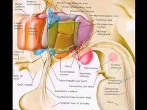
Limbic system

Limbic system (or Paleomammalian brain) is a set of brain structures including the hippocampus, amygdala, anterior thalamic nuclei, and limbic cortex, which support a variety of functions including emotion, behavior, long term memory, and olfaction.
Essentially the limbic system is the set of brain structures that forms the inner border of the cortex. In an abstract topological sense, each cortical hemisphere can be thought of as a sphere of gray matter, with a hole punched through it in the area where nerve fibers connect it to the subcortical structures of the basal forebrain. The hole is surrounded by a ring of cortical and noncortical areas that combine to make up the limbic system. The cortical components generally have fewer layers than the classical 6-layered neocortex, and are often classified as allocortex or archicortex.
The limbic system includes many structures in the cerebral cortex and sub-cortex of the brain. The term has been used within psychiatry and neurology, although its exact role and definition has been revised considerably since the term was introduced. The following structures are, or have been considered to be, part of the limbic system:
- Amygdala: Involved in signaling the cortex of motivationally significant stimuli such as those related to reward and fear in addition to social functions such as mating.
- Hippocampus:Required for the formation of long-term memories and implicated in maintenance of cognitive maps for navigation.
- Parahippocampal gyrus: Plays a role in the formation of spatial memory * Cingulate gyrus:Autonomic functions regulating heart rate, blood pressure and cognitive and attentional processing
- Fornix:carries signals from the hippocampus to the mammillary bodies and septal nuclei.
- Hypothalamus:Regulates the autonomic nervous system via hormone production and release. Affects and regulates blood pressure, heart rate, hunger, thirst, sexual arousal, and the sleep/wake cycle
- Thalamus: The "relay station" to the cerebral cortex
- Mammillary body: Important for the formation of memory
- Pituitary gland: secretes hormones regulating homeostasis
- Dentate gyrus: thought to contribute to new memories and to regulate happiness.
- Entorhinal cortex and piriform cortex: Receive smell input in the olfactory system.
- Fornicate gyrus: Region encompassing the cingulate, hippocampus, and parahippocampal gyrus
- Olfactory bulb: Olfactory sensory input
- Nucleus accumbens: Involved in reward, pleasure, and addiction
- Orbitofrontal cortex: Required for decision making
Function The limbic system operates by influencing the endocrine system and the autonomic nervous system. It is highly interconnected with the nucleus accumbens, the brain's pleasure center, which plays a role in sexual arousal and the "high" derived from certain recreational drugs. These responses are heavily modulated by dopaminergic projections from the limbic system. In 1954, Olds and Milner found that rats with metal electrodes implanted into their nucleus accumbens repeatedly pressed a lever activating this region, and did so in preference to eating and drinking, eventually dying of exhaustion.
The limbic system is also tightly connected to the prefrontal cortex. Some scientists contend that this connection is related to the pleasure obtained from solving problems. To cure severe emotional disorders, this connection was sometimes surgically severed, a procedure of psychosurgery, called a prefrontal lobotomy (this is actually a misnomer). Patients who underwent this procedure often became passive and lacked all motivation.
Nonspecific Defenses-Interferon

Interferons (IFNs) are natural cell-signaling proteins produced by the cells of the immune system of most vertebrates in response to challenges such as viruses, parasites and tumor cells. Interferons belong to the large class of glycoproteins known as cytokines. Interferons are produced by a wide variety of cells in response to the presence of double-stranded RNA, a key indicator of viral infection. Interferons assist the immune response by inhibiting viral replication within host cells, activating natural killer cells and macrophages, increasing antigen presentation to lymphocytes, and inducing the resistance of host cells to viral infection.
Types of interferon
There are three major classes of interferons that have been described for humans according to the type of receptor through which they signal:
- Interferon type I: All type I IFNs bind to a specific cell surface receptor complex known as the IFN-α receptor (IFNAR) that consists of IFNAR1 and IFNAR2 chains. The type I interferons present in humans are IFN-α, IFN-β and IFN-ω.
- Interferon type II: Binds to IFNGR. In humans this is IFN-γ.
- Interferon type III: Signal through a receptor complex consisting of IL10R2 (also called CRF2-4) and IFNLR1 (also called CRF2-12). Interferon type III is not accepted as a separate classification in mainstream Medicine.
Interferons in general have several effects in common. They are antiviral and possess antioncogenic properties, macrophage and natural killer cell activation, and enhancement of major histocompatibility complex glycoprotein classes I and II, and thus presentation of foreign (microbial) peptides to T cells. In a majority of cases, the production of interferons is induced in response to microbes such as viruses and bacteria and their products (viral glycoproteins, viral RNA, bacterial endotoxin, bacterial flagella, CpG sites), as well as mitogens and other cytokines, for example interleukin 1, interleukin 2, interleukin-12, tumor necrosis factor and colony-stimulating factor, that are synthesised in the response to the appearance of various antigens in the body. Their metabolism and excretion take place mainly in the liver and kidneys. They rarely pass the placenta but they can cross the blood-brain barrier.
The therapeutically used forms are denoted by Greek letters indicating their origin: leukocytes, fibroblasts, and lymphocytes for interferon-alpha, -beta and -gamma, respectively.
Nonspecific Defenses- Fever

Fever is a frequent medical sign that describes an increase in internal body temperature to levels above normal.Fever is caused by an elevation in the thermoregulatory set-point, causing typical body temperature (generally and problematically considered to be 37 °C ±1 °C, or approximately 99 ±2 °F; see below for specifics) to rise, and effector mechanisms are enacted as a result. A feverish individual has a general feeling of cold despite an increased body temperature, and increases in heart rate, muscle tone and shivering, all of which are caused by the body's attempts to counteract the newly-perceived hypothermia and reach the new thermoregulatory set-point.
Fever is considered one of the body's immune mechanisms to attempt a neutralization of a perceived threat inside the body, be it bacterial or viral. Carl Wunderlich discovered that fever is not a disease, but the body's response to a disease.
Transport of Oxygen

In the lungs, oxygen diffuses from alveolar air into the blood because the venous blood has a lower partial pressure. The oxygen dissolves in the blood. Only a small amount is carried as a physical solution (0.31 ml per 100 ml). The remainder of the oxygen is carried in chemical combination with the hemoglobin in red blood cells (erthrocytes).
Hemoglobin (molecular weight of 68,000) is made from 4 hemes, a porphyrin ring containing iron and globin, a 4 protein chains. Oxygen is bound to the iron for the transport process. Hemoglobin (HHgb) behaves as a weak acid (K = 1.4 x 10⁻⁸; pKa = 7.85). Oxyhemoglobin (HHgbO2) also behaves as a weak acid (K = 2.5 x 10⁻⁷; pKa = 6.6).
Because both forms of hemoglobin are weak acids, and a relationship of the numerical values of the equilibrium constants, the net reaction for the interaction of oxygen with hemoglobin results in the following equilibrium:
HHgb + O 2 <===> HgbO 2 + H+If 2 is increased in the blood at the lungs, the equilibrium shifts to the right and H+ ions increase.
Oxyhemoglobin can be caused to release oxygen by the addition of H+ ions at the cells. The difference in pH (7.44) of arterial blood and venous blood (pH = 7.35) is sufficient to cause release of oxygen from hemoglobin at the tissue cells.
The Immune Mechanism

Immune mechanism is designed to protect body against invading microorganisms and foreign potentially harmful molecules.There are four types of immune mechanism; the nature of invading antigen determines which type of mechanism bought into action.Certain antigen promotes exaggerated response called hypersensitive reaction or an allergy, which may be harmful to body tissues.
Type 1 reaction is allergic response to foreign substances usually protein entering the body it is a immediate reaction occurs within the minutes or hours of antigen entering the body.In type 1 response the antigen enters the body and stimulate B-lymphocyte to produce antibodies. These antibodies adhere Mast cells in the vessel wall, they neutralizes the antigen and the Mast cell releases the chemical which causes streaming eyes, sneezing symptomatic fever.
In type 2 reactions is initiated by antigens, which are part of tissue cell. Antigen enters the blood stream and invokes the production of antibodies.Antibodies destroy the antigens, but also cause cross reaction with the blood cell which can lead to cell damage, example Mismatch Blood transfusion in which antibodies are formed against donor red cells, which leads to their destruction.
In type 3 reactions is a immediate reaction occurring within few hours of antigenic stimulation. Antigen enters the blood stream, which already filled with antibodies formed during the previous exposure with same antigens.Antibodies form a complex with antigen a blood protein called complement. The complex may damage tissue such as glomeruli in kidney by blocking the capillaries.
Type 4 Reaction is a delayed immune response, which occurs more than 24 hours after the initial contact with the antigen. The antigens enter the blood stream, where it stimulate T lymphocytes to produce antibodies, which remain attached to the cell wall, the anitbodies, and then destroys the antigens.Once T-lymphocytes are been sensitized by antigens the can produce antibodies and confer immunity
Continuous Circuit

In an adult's body about ten pints of blood are continuously being pumped by the heart through sixty thousand miles of blood vessels. Blood, which has acquired oxygen in the lungs, leaves the heart through the aorta, an inch-wide tube. The aorta branches into large arteries, which take the blood to the neck, head and arms. It then passes down the middle of the body carrying blood to the kidneys, liver, intestines and legs. The large arteries further divide into smaller arterioles and, then finally, into minute capillaries, which surround the body cells. Blood then passes into venules, small veins, and then into larger veins, which feed into the superior vena cava and the inferior vena cava, which return the deoxygenated blood to the heart and then to the lung, where the blood gives up carbon dioxide and acquires a fresh supply of oxygen. >
Red Blood Cells
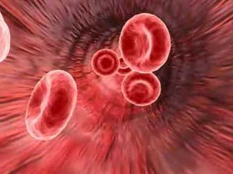
Red blood cells are the most common type of blood cell and the vertebrate body's principal means of delivering oxygen to the body tissues via the blood. They take up oxygen in the lungs or gills and release it while squeezing through the body's capillaries. The cells are filled with hemoglobin, a biomolecule that can bind to oxygen. The blood's red color is due to the color of hemoglobin. In humans, red blood cells develop in the bone marrow and live for about 120 days; they take the form of flexible biconcave disks that lack a cell nucleus and organelles and they cannot synthesize protein.
Red blood cells are also known as RBCs, red blood corpuscles (an archaic term), haematids or erythrocytes (from Greek erythros for "red" and kytos for "hollow", with cyte translated as "cell" in modern usage). The capitalized term Red Blood Cells is the proper name in the US for erythrocytes in storage solution used in transfusion medicine.
Laser generation of nanoparticles in liquids

An invisible (infrared) pulsed laser beam transmits a liquid (water) and is ablating a solid (gold) releasing stable nanoparticles (positively charged gold nanoparticle with negative solvatisation zeta potential) .

Toll-like receptor

Toll-like receptors (TLRs) are a class of proteins that play a key role in the innate immune system. They are single membrane-spanning non-catalytic receptors that recognize structurally conserved molecules derived from microbes. Once these microbes have breached physical barriers such as the skin or intestinal tract mucosa, they are recognized by TLRs which activates immune cell responses.
They receive their name from their similarity to the protein coded by the Toll gene identified in Drosophila in 1985 by Christiane Nüsslein-Volhard.
Diversity
TLRs are a type of pattern recognition receptor (PRR) and recognize molecules that are broadly shared by pathogens but distinguishable from host molecules, collectively referred to as pathogen-associated molecular patterns (PAMPs). TLRs together with the Interleukin-1 receptors form a receptor superfamily, known as the "Interleukin-1 Receptor/Toll-Like Receptor Superfamily"; all members of this family have in common a so-called TIR (Toll-IL-1 receptor) domain.
Three subgroups of TIR domains exist. Proteins with subgroup 1 TIR domains are receptors for interleukins that are produced by macrophages, monocytes and dendritic cells and all have extracellular Immunoglobulin (Ig) domains. Proteins with subgroup 2 TIR domains are classical TLRs, and bind directly or indirectly to molecules of microbial origin. A third subgroup of proteins containing TIR domains consists of adaptor proteins that are exclusively cytosolic and mediate signaling from proteins of subgroups 1 and 2.
TLRs are present in vertebrates, as well as in invertebrates. Molecular building blocks of the TLRs are represented in bacteria and in plants, and in the latter kingdom, are well known to be required for host defence against infection. The TLRs thus appear to be one of the most ancient, conserved components of the immune system.
Discovery
Toll-like receptors are now counted among the key molecules that alert the immune system to the presence of microbial infections. They are named for their similarity to Toll, a receptor first identified in the fruit fly Drosophila melanogaster, and originally known for its developmental function in that organism. In 1996, Toll was found by Jules A. Hoffmann and his colleagues to have an essential role in the fly's immunity to fungal infection,which it achieved by activating the synthesis of antimicrobial peptides.
The first reported human Toll-like receptor was described by Nomura and colleagues in 1994, mapped to a chromosome by Taguchi and colleagues in 1996.Because the immune function of Toll in Drosophila was not then known, it was assumed that TIL (now known as TLR1) might participate in mammalian development. However, in 1991 (prior to the discovery of TIL) it was observed that a molecule with a clear role in immune function in mammals, the interleukin-1 (IL-1) receptor, also had homology to drosophila Toll; the cytoplasmic portions of both molecules were similar.
In 1997, Charles Janeway and Ruslan Medzhitov showed that a Toll-like receptor now known as TLR4 could, when artificially ligated using antibodies, induce the activation of certain genes necessary for initiating an adaptive immune response. However, the function of the TLRs remained unknown in the wake of this work, and in particular, no ligand had been identified for any mammalian TLR.
TLR function was discovered by Bruce A. Beutler and colleagues.These workers used positional cloning to prove that mice that could not respond to LPS had mutations that abolished the function of TLR4. This identified TLR4 as a key component of the receptor for LPS, and strongly suggested that other Toll-like receptors might detect other signature molecules of microbes, such as those mentioned above.
In turn, the other TLR genes were ablated in mice by gene targeting, largely in the laboratory of Shizuo Akira and colleagues. Each TLR is now believed to detect a discrete collection of molecules of microbial origin, and to signal the presence of infections.
Ligands
Because the specificity of Toll-like receptors (and other innate immune receptors) cannot easily be changed in the course of evolution, these receptors recognize molecules that are constantly associated with threats (i.e. pathogen or cell stress) and are highly specific to these threats (i.e. cannot be mistaken for self molecules). Pathogen-associated molecules that meet this requirement are usually critical to the pathogen's function and cannot be eliminated or changed through mutation; they are said to be evolutionarily conserved. Well conserved features in pathogens include bacterial cell-surface lipopolysaccharides (LPS), lipoproteins, lipopeptides and lipoarabinomannan; proteins such as flagellin from bacterial flagella; double-stranded RNA of viruses or the unmethylated CpG islands of bacterial and viral DNA; and certain other RNA and DNA. For most of the TLRs, ligand recognition specificity has now been established by gene targeting (also known as "gene knockout"): a technique by which individual genes may be selectively deleted in mice.
Endogenous ligands
The stereotypic inflammatory response provoked by TLR activation has prompted speculation that endogenous activators of TLRs might participate in autoimmune diseases. TLRs have been suspected of binding to host molecules including fibrinogen (involved in blood clotting) and heat shock proteins (HSPs)and host DNA.
Signaling
TLRs are believed to function as dimers. Though most TLRs appear to function as homodimers, TLR2 forms heterodimers with TLR1 or TLR6, each dimer having a different ligand specificity. TLRs may also depend on other co-receptors for full ligand sensitivity, such as in the case of TLR4's recognition of LPS, which requires MD-2. CD14 and LPS Binding Protein (LBP) are known to facilitate the presentation of LPS to MD-2.
The adapter proteins and kinases that mediate TLR signaling have also been targeted. In addition, random germline mutagenesis with ENU has been used to decipher the TLR signaling pathways. When activated, TLRs recruit adapter molecules within the cytoplasm of cells in order to propagate a signal. Four adapter molecules are known to be involved in signaling. These proteins are known as MyD88, Tirap (also called Mal), Trif, and Tram. The adapters activate other molecules within the cell, including certain protein kinases (IRAK1, IRAK4, TBK1, and IKKi) that amplify the signal, and ultimately lead to the induction or suppression of genes that orchestrate the inflammatory response. In all, thousands of genes are activated by TLR signaling, and collectively, the TLRs constitutes one of the most pleiotropic yet tightly regulated gateways for gene modulation.
Activation and effects
Following activation by ligands of microbial origin, several reactions are possible. Immune cells can produce signalling factors called cytokines which trigger inflammation. In the case of a bacterial factor, the pathogen might be phagocytosed and digested, and its antigens presented to CD4+ T cells. In the case of a viral factor, the infected cell may shut off its protein synthesis and may undergo programmed cell death (apoptosis). Immune cells that have detected a virus may also release anti-viral factors such as interferons.
The discovery of the Toll-like receptors finally identified the innate immune receptors that were responsible for many of the innate immune functions that had been studied for many years. Interestingly, TLRs seem only to be involved in the cytokine production and cellular activation in response to microbes, and do not play a significant role in the adhesion and phagocytosis of microorganisms.
Drugs interactions
Imiquimod (cardinally used in dermatology), and its successor R848, are ligands for TLR7 and TLR8
Toll-like receptor. (2009, May 4). In Wikipedia, The Free Encyclopedia. Retrieved 13:36, May 12, 2009, from http://en.wikipedia.org/w/index.php?title=Toll-like_receptor&oldid=287743888
Toll-like receptor. (2009, May 4). In Wikipedia, The Free Encyclopedia. Retrieved 13:36, May 12, 2009, from http://en.wikipedia.org/w/index.php?title=Toll-like_receptor&oldid=287743888
Heartbeat

Every single 'beat' of the heart involves five major stages: First, "Late diastole" which is when the semilunar valves close, the Av valves open and the whole heart is relaxed. Second, "Atrial systole" when atria is contracting, AV valves open and blood flows from atrium to the ventricle. Third, "Isovolumic ventricular contraction" it is when the ventricles begin to contract, AV valves close, as well as the semilunar valves and there is no change in volume. Fourth, "ventricular ejection", Ventricles are empty, they are still contracting and the semilunar valves are open.

The fifth stage is: "Isovolumic ventricular relaxation", Pressure decreases, no blood is entering the ventricles, ventricles stop contracting and begin to relax, semilunars are shut because blood in the aorta is pushing them shut. Throughout the cardiac cycle, the blood pressure increases and decreases. The cardiac cycle is coordinated by a series of electrical impulses that are produced by specialized heart cells found within the sino-atrial node and the atrioventricular node. The cardiac muscle is composed of myocytes which initiate their own contraction without help of external nerves
The fifth stage is: "Isovolumic ventricular relaxation", Pressure decreases, no blood is entering the ventricles, ventricles stop contracting and begin to relax, semilunars are shut because blood in the aorta is pushing them shut. Throughout the cardiac cycle, the blood pressure increases and decreases. The cardiac cycle is coordinated by a series of electrical impulses that are produced by specialized heart cells found within the sino-atrial node and the atrioventricular node. The cardiac muscle is composed of myocytes which initiate their own contraction without help of external nerves
Atrial systole
Atrial systole is the contraction of the heart muscle (myocardia) of the left and right atria. Normally, both atria contract at the same time. The term systole is synonymous with contraction (movement or shortening) of a muscle. Electrical systole is the electrical activity that stimulates the myocardium of the chambers of the heart to make them contract. This is soon followed by Mechanical systole, which is the mechanical contraction of the heart.
As the atria contract, the blood pressure in each atrium increases, forcing additional blood into the ventricles. The additional flow of blood is called atrial kick.
70% of the blood flows passively down to the ventricles, so the atria do not have to contract a great amount.
Atrial kick is absent if there is loss of normal electrical conduction in the heart, such as during atrial fibrillation, atrial flutter, and complete heart block. Atrial kick is also different in character depending on the condition of the heart, such as stiff heart, which is found in patients with diastolic dysfunction.
Detection of atrial systole
Electrical systole of the atria begins with the onset of the P wave on the ECG. The wave of bipolarization (or depolarization) that stimulates both atria to contract at the same time is due to sinoatrial node which is located on the upper wall of the right atrium. 30% of the ventricles are filled during this phase.
Ventricular systole
Ventricular systole is the contraction of the muscles (myocardia) of the left and right ventricles.At the later part of the ejection phase, although the ventricular pressure falls below the aortic pressure, the aortic valve remains patent because of the inertial energy of the ejected blood.
The graph of aortic pressure throughout the cardiac cycle displays a small dip which coincides with the aortic valve closure. The dip in the graph is immediately followed by a brief rise then gradual decline. The small rise in the graph is known as the "dicrotic notch" or "incisure", and represents a transient increase in aortic pressure. Just as the ventricles enter into diastole, the brief reversal of flow from the aorta back into the left ventricle causes the aortic valves to shut. This results in the slight increase in aortic pressure caused by the elastic recoil of the semilunar valves and aorta.

Pituitary Gland

A small oval endocrine gland attached to the base of the vertebrate brain and consisting of an anterior and a posterior lobe, the secretions of which control the other endocrine glands and influence growth, metabolism, and maturation. Also called hypophysis, pituitary body.
It is most structurally and functionally complex organ of endocrine system. All vertebrates have a pituitary gland with a common basic structure and function. In addition to its endocrine functions, the pituitary may play a role in the immune response.
It is most structurally and functionally complex organ of endocrine system. All vertebrates have a pituitary gland with a common basic structure and function. In addition to its endocrine functions, the pituitary may play a role in the immune response.
The hypophysis of all vertebrates has two major segments—the neurohypophysis (a neural component) and the adenohypophysis (an epithelial component)—each with a different embryological origin. The neurohypophysis develops from a downward process of the diencephalon (the base of the brain), whereas the adenohypophysis originates as an outpocketing of the primitive buccal epithelium, known as Rathke's pouch. The adenohypophysis has three distinct subdivisions: the pars tuberalis, the pars distalis, and the pars intermedia. The neurohypophysis comprises the pars nervosa and the infundibulum. The latter consists of the infundibular stalk and the median eminence of the tuber cinereum.
The anterior pituitaryIt contains five different types of cell, each of which produce one particular hormone, with the exception of the ‘gonadotrophs’ which produce two: namely luteinizing hormone (LH) and follicular stimulating hormone (FSH). All the hormones are peptide or protein in nature, varying in size from 39 amino acids (ACTH) to 204 amino acids (LH and FSH). The hormones fall into two groups: the first contains the four trophic hormones (from the Greek for nourishment), which control other endocrine glands; the second contains prolactin and growth hormone, which have more widespread effects in the body.
The trophic hormones act to stimulate secretion of hormone from the target gland and to maintain its function and, if present in high concentrations, will cause the gland to enlarge. They are:
- thyroid stimulating hormone (TSH), which stimulates the secretion of the thyroid hormones;
- adrenocorticotrophic hormone (ACTH), which acts on the adrenal cortex to promote the release of cortisol;
- gonadotrophins LH and FSH, which act on the ovaries and testes. They are however named after their effects in women; FSH stimulates growth of the ovarian follicle containing the ovum or egg and LH stimulates production of oestrogen and progesterone from the ovary. The actions in the male are analogous; FSH stimulates sperm production and LH stimulates testosterone production by the testes.
Prolactin acts chiefly to cause milk production in the breasts.
Growth hormone has widespread effects, necessary not only for growth itself but also for metabolism throughout life.
Because the pituitary controls so many endocrine functions in the body it has been called ‘the conductor of the endocrine orchestra’, but more recent discoveries suggested that this term more properly belongs to the hypothalamus, with the pituitary being comparable to the leader of the orchestra. Since the nerves going to the anterior pituitary only supply the blood vessels there was some debate as to how the gland was controlled. It is now known that the hypothalamus produces stimulatory and inhibitory hormones, and that these reach the anterior pituitary via a network of small blood vessels or capillaries. The hormones are produced in nerve cells whose endings abut on the capillaries at the top of the pituitary stalk. This control of the pituitary by the central nervous system allows blood concentrations of the hormones to respond to a variety of external stimuli including stress. It also allows for complex patterning of release. Pituitary hormones in general are released in a pulsatile fashion, with many pulses during the day, and they can also show 24 hour (diurnal) rhythms. The gonadotrophins, linked into the human menstrual cycle, show a 28 day rhythm, while in animals which are seasonal breeders prolactin shows a seasonal rhythm. Blood concentrations of pituitary hormones are controlled not only by the hypothalamic hormones but by feedback, usually negative, exerted by target organ hormones such as cortisol or progesterone.
The posterior pituitary
Two hormones are released from the posterior lobe, oxytocin and vasopressin (syn. antidiuretic hormone) . These, like the releasing hormones that reach the anterior lobe, are produced within nerve cells in the hypothalamus. But in this case the axons travel right down the pituitary stalk, and the nerve endings release the hormones directly into the bloodstream (see endocrine). The activity of the posterior pituitary hormones was established around 1900 in the UK by Schafer (a physiologist) and his colleagues working on what proved to be the actions of vasopressin, and Dale, a pharmacologist and Nobel Prize winner working on oxytocin. Vasopressin plays a role in water balance and the maintenance of blood pressure, normal circulating concentrations causing water to be retained by the kidney and higher concentrations causing blood vessels to constrict, thus raising blood pressure. As with the anterior pituitary, control via the hypothalamus means that release of posterior pituitary hormones can be regulated by a variety of nervous inputs; the main stimuli for vasopressin release are an increase in the concentration of the blood plasma and a decrease in circulating blood volume, both of which reflect a fall in total body water. Oxytocin is important for the birth of an infant and for delivery of the milk supply.
Actin Polymerization

Actin is the component of the cytoskeletal system that allows movement of cells and cellular processes. It works in conjunction or in tandem with other components of the system. Like the other components, it can undergo constant rearrangement to produce movement. Actin filaments are also called microfilaments, or "thin filaments" to distinguish them from intermediate filaments.
During the polymerization process, adenosine 5'-triphosphate (ATP) that is bound to G-actin is hydrolyzed to adenosine 5'-diphosphate (ADP) that is bound to F-actin. The hydrolysis reaction occurs on the F-actin subsequent to the polymerization reaction in two steps: cleavage of ATP followed by the slower release of inorganic phosphate (Pi). As a result, at high rates of filament growth a transient cap of ATP-actin subunits exists at the ends of elongating filaments, and at steady state a stabilizing cap of ADP.Pi-actin subunits exists at the barbed ends of filaments. Cleavage of ATP results in a highly stable filament with bound ADP.Pi, and release of Pi destabilizes the filament. Thus these two steps of the hydrolytic reaction provide potential mechanisms for regulating the monomer-polymer transition.
Pyrosequencing

Pyrosequencing is a method of DNA sequencing (determining the order of nucleotides in DNA) based on the "sequencing by synthesis" principle, which relies on detection of pyrophosphate release on nucleotide incorporation rather than chain termination with dideoxynucleotides.The technique was developed by Pål Nyrén and his student Mostafa Ronaghi at the Royal Institute of Technology in Stockholm in 1996.
"Sequencing by synthesis" involves taking a single strand of the DNA to be sequenced and then synthesizing its complementary strand enzymatically. The Pyrosequencing method is based on detecting the activity of DNA polymerase (a DNA synthesizing enzyme) with another chemiluminescent enzyme. Essentially, the method allows sequencing of a single strand of DNA by synthesizing the complementary strand along it, one base pair at a time, and detecting which base was actually added at each step. The template DNA is immobilized, and solutions of A, C, G, and T nucleotides are added and removed after the reaction, sequentially. Light is produced only when the nucleotide solution complements the first unpaired base of the template. The sequence of solutions which produce chemiluminescent signals allows the determination of the sequence of the template.
ssDNA template is hybridized to a sequencing primer and incubated with the enzymes DNA polymerase, ATP sulfurylase, luciferase and apyrase, and with the substrates adenosine 5´ phosphosulfate (APS) and luciferin.
1. The addition of one of the four deoxynucleotide triphosphates (dNTPs)(in the case of dATP we add dATPαS which is not a substrate for a luciferase) initiates the second step. DNA polymerase incorporates the correct, complementary dNTPs onto the template. This incorporation releases pyrophosphate (PPi) stoichiometrically.
2. ATP sulfurylase quantitatively converts PPi to ATP in the presence of adenosine 5´ phosphosulfate. This ATP acts as fuel to the luciferase-mediated conversion of luciferin to oxyluciferin that generates visible light in amounts that are proportional to the amount of ATP. The light produced in the luciferase-catalyzed reaction is detected by a camera and analyzed in a program.
3. Unincorporated nucleotides and ATP are degraded by the apyrase, and the reaction can restart with another nucleotide.
Currently, a limitation of the method is that the lengths of individual reads of DNA sequence are in the neighborhood of 300-500 nucleotides, shorter than the 800-1000 obtainable with chain termination methods (e.g. Sanger sequencing). This can make the process of genome assembly more difficult, particularly for sequence containing a large amount of repetitive DNA. As of 2007, pyrosequencing is most commonly used for resequencing or sequencing of genomes for which the sequence of a close relative is already available.
The templates for pyrosequencing can be made both by solid phase template preparation (Streptavidin coated magnetic beads) and enzymatic template preparation (Apyrase+Exonuclease).


