Pages

Showing posts with label brain. Show all posts

Olfactory Pathway Animation

The olfactory system is the sensory system used for olfaction. Most mammals and reptiles have two distinct parts to their olfactory system: a main olfactory system and an accessory olfactory system. The main olfactory system detects volatile, airborn substances, while the accessory olfactory system senses fluid-phase stimuli. Behavioral evidence indicates that most often, the stimuli detected by the accessory olfactory system are pheromones.


Bookmark and Share 

The olfactory system is often spoken of along with the gustatory system as the chemosensory senses because both transduce chemical signals into perception.

The mechanism of the olfactory system can be divided into a peripheral one, sensing an external stimulus and encoding it as an electric signal in neurons, and a central one, where all signals are integrated and processed in the central nervous system.


In mammals, the main olfactory system detects odorants that are inhaled through the nose, where they contact the main olfactory epithelium, which contains various olfactory receptors. These can distinguish a new odor from the background environmental odors and determine the concentration of the odor.

These olfactory receptors are connected to olfactory receptor neurons in the olfactory epithelium, which transduce receptoractivation into electrical signals in neurons. The signals travel along the olfactory nerve, which belongs to the peripheral nervous system. This nerve terminates in the olfactory bulb, which belongs to the central nervous system.

Nature video on how brain sees


At the micro-scale the brain is a mess; a thick tangle of nerve cells connected at synapses. Mapping just a tiny portion of this mess, a few hundred cells, is a huge challenge. You have to wonder if it's worth the effort. But seeing exactly how brain cells are wired together is giving us new insights into brain function. The researchers who made the 3D maps in this video discovered a new type of cell and worked out how insects see movement. If you've ever tried to swat a fly you'll know how good they are at sensing motion!

Schizophrenia


Schizophrenia is a chronic, severe, and disabling brain disorder that has been recognized throughout recorded history. It affects about 1 percent of Americans.
People with schizophrenia may hear voices other people don't hear or they may believe that others are reading their minds, controlling their thoughts, or plotting to harm them. These experiences are terrifying and can cause fearfulness, withdrawal, or extreme agitation. People with schizophrenia may not make sense when they talk, may sit for hours without moving or talking much, or may seem perfectly fine until they talk about what they are really thinking. Because many people with schizophrenia have difficulty holding a job or caring for themselves, the burden on their families and society is significant as well.

Bookmark and Share  Subscribe in a reader

Available treatments can relieve many of the disorder's symptoms, but most people who have schizophrenia must cope with some residual symptoms as long as they live. Nevertheless, this is a time of hope for people with schizophrenia and their families. Many people with the disorder now lead rewarding and meaningful lives in their communities. Researchers are developing more effective medications and using new research tools to understand the causes of schizophrenia and to find ways to prevent and treat it.

This brochure presents information on the symptoms of schizophrenia, when the symptoms appear, how the disease develops, current treatments, support for patients and their loved ones, and new directions in research.


Psychotic symptoms (such as hallucinations and delusions) usually emerge in men in their late teens and early 20s and in women in their mid-20s to early 30s. They seldom occur after age 45 and only rarely before puberty, although cases of schizophrenia in children as young as 5 have been reported. In adolescents, the first signs can include a change of friends, a drop in grades, sleep problems, and irritability. Because many normal adolescents exhibit these behaviors as well, a diagnosis can be difficult to make at this stage. In young people who go on to develop the disease, this is called the "prodromal" period.

Research has shown that schizophrenia affects men and women equally and occurs at similar rates in all ethnic groups around the world.

Neuron

The complexity and diversity in nervous systems is dependent on the interconnections between neurons, which rely on a limited number of different signals transmitted within the neurons to other neurons or to muscles and glands. The signals are produced and propagated by chemical ions that produce an electrical charge that moves along the neuron.

  Subscribe in a reader
Neurons exist in a number of different shapes and sizes and can be classified by their morphology and function. The anatomist Camillo Golgi grouped neurons into two types; type I with long axons used to move signals over long distances and type II without axons. type I cells can be further divided by where the cell body or soma is located. The basic morphology of type I neurons, represented by spinal motor neurons, consists of a cell body called the soma and a long thin axon which is covered by the myelin sheath. Around the cell body is a branching dendritic tree that receives signals from other neurons. The end of the axon has branching terminals (axon terminal) that release transmitter substances into a gap called the synaptic cleft between the terminals and the dendrites of the next neuron.
The anatomy and the properties of the surface membrane determine the behavior of a neuron. The surface membrane is not uniform over the entire length of a neuron, but is modified in specific areas: some regions secrete transmitter substances while other areas respond to the transmitter. Other areas of the neuron membrane have passive electrical properties that effect capacitance and resistance. Within the neuron membrane there are gated ion channels that vary in type, including fast response sodium channels that are voltage-gated and are used to send rapid signals.
Neurons communicate by chemical and electrical synapses in a process known as synaptic transmission. The fundamental process that triggers synaptic transmission is the action potential, a propagating electrical signal that is generated by exploiting the electrically excitable membrane of the neuron. This is also known as a wave of depolarization.
Fully differentiated neurons are permanently amitotic; however, recent research shows that additional neurons throughout the brain can originate from neural stem cells found in high concentrations in (but throughout the brain) the subventricular zone and subgranular zone through the process of neurogenesis.

Concussions animation

Every year, millions of people in the U.S. sustain head and brain injuries. Some are minor because the skull is quite good at protecting the brain. More than half are bad enough that people must go to the hospital. Serious head injuries can lead to permanent brain damage or death.

Symptoms of minor head injuries usually go away without treatment. Serious head injuries need emergency treatment. Clues that a head injury may be serious include

Aneurysms Animation

A cerebral aneurysm (also known as an intracranial or intracerebral aneurysm) is a weak or thin spot on a blood vessel in the brain that balloons out and fills with blood. The bulging aneurysm can put pressure on a nerve or surrounding brain tissue. It may also leak or rupture, spilling blood into the surrounding tissue (called a hemorrhage). Some cerebral aneurysms, particularly those that are very small, do not bleed or cause other problems. Cerebral aneurysms can occur anywhere in the brain, but most are located along a loop of arteries that run between the underside of the brain and the base of the skull.

  Subscribe in a reader

Most cerebral aneurysms are congenital, resulting from an inborn abnormality in an artery wall. Cerebral aneurysms are also more common in people with certain genetic diseases, such as connective tissue disorders and polycystic kidney disease, and certain circulatory disorders, such as arteriovenous malformations.


Other causes include trauma or injury to the head, high blood pressure, infection, tumors, atherosclerosis (a blood vessel disease in which fats build up on the inside of artery walls) and other diseases of the vascular system, cigarette smoking, and drug abuse. Some investigators have speculated that oral contraceptives may increase the risk of developing aneurysms.

Aneurysms that result from an infection in the arterial wall are called mycotic aneurysms. Cancer-related aneurysms are often associated with primary or metastatic tumors of the head and neck. Drug abuse, particularly the habitual use of cocaine, can inflame blood vessels and lead to the development of brain aneurysms.


Text Source: NINDS

Human Suprachiasmatic Nucleus

The suprachiasmatic nucleus (SCN) is a bilateral region of the brain, located in the hypothalamus, that is responsible for controlling endogenous circadian rhythms. The neuronal and hormonal activities it generates regulate many different body functions over a 24-hour period.
The SCN contains several cell types and several different peptides (including vasopressin and vasoactive intestinal peptide) and neurotransmitters, and interacts with many other regions of the brain.
The SCN is situated in the anterior hypothalamus immediately dorsal to the optic chiasm (CHO) bilateral to (on either side of) the third ventricle.


Bookmark and Share  Subscribe in a reader

Circadian effects
The SCN receives inputs from specialized photoreceptive retinal ganglion cells, via the retinohypothalamic tract.
Destruction of the SCN leads to a complete loss of circadian rhythm. Rats with damage to the SCN have no circadian rhythms, i.e., they sleep the same total amount, but polyphasically for random lengths at a time.
The SCN also controls 'slave oscillators' in the peripheral tissues, which exhibit their own ~24 hour rhythms, but are crucially synchronized by the SCN.
The importance of entraining our bodies to an exogenous cue, such as daylight, is reflected by several circadian rhythm sleep disorders, where this process does not function normally.
Neurons in the ventrolateral SCN (vlSCN) have the ability for light-induced gene expression. If light is turned on at night, the vlSCN relays this information throughout the SCN, in a process called entrainment.

Neurons in the dorsomedial SCN (dmSCN) are believed to make an endogenous 24-hour rhythm that can persist under constant darkness (in humans averaging about 24h 11min). Melanopsin-containing ganglion cells in the retina have a direct connection to the SCN via the retinohypothalamic tract.
The SCN sends information to other hypothalamic nuclei and the pineal gland to modulate body temperature and production of hormones such as cortisol and melatonin.
Other signals from the retina
  • The SCN is one of four nuclei that receive nerve signals directly from the retina.
  • The other three are the lateral geniculate nucleus (LGN), the superior colliculus, and the pretectum:
  • The LGN passes information about color, contrast, shape, and movement on to the visual cortex and itself signals to the SCN.
  • The superior colliculus controls the movement and orientation of the eyeball.
  • The pretectum controls the size of the pupil.
Gene expression
The circadian rhythm in the SCN is generated by a gene expression cycle in individual SCN neurons. This cycle has been well conserved through evolution, and is essentially similar in cells from many widely different organisms that show circadian rhythms.
Electrophysiology
Neurons in the SCN fire action potentials in a 24-hour rhythm. At mid-day, the firing rate reaches a maximum, and, during the night, it falls again. How the gene expression cycle (so-called the core clock) connects to the neural firing remains unknown.
Many SCN neurons are sensitive to light stimulation via the retina, and sustainedly firing action potentials during a light pulse (~30 seconds) in rodents. The photic response is likely linked to effects of light on circadian rhythms. In addition, focal application of melatonin can decrease firing activity of these neurons, suggesting that melatonin receptors present in the SCN mediate phase-shifting effects through the SCN.

Building the Brain: From Simplicity to Complexity

What are the mechanisms by which neurons differentiate to achieve the spectacular complexity of the brain? Join UCSD's Nick Spitzer as he explains what we know about this process


Spatial memory

Spatial memory is the part of memory responsible for recording information about one's environment and its spatial orientation. For example, a person's spatial memory is required in order to navigate around a familiar city, just as a rat's spatial memory is needed to learn the location of food at the end of a maze. It is often argued that a person's, or an animal's, spatial memories are summarised in a cognitive map.

Spatial memories are formed after an organism gathers and processes sensory information about its surroundings (especially vision and proprioception). In general, mammals require a functioning hippocampus (particularly area CA1) in order to form and process memories about space. There is some evidence that human spatial memory is strongly tied to the right hemisphere of the brain.


Spatial learning requires both NMDA and AMPA receptors, consolidation requires NMDA receptors, and the retrieval of spatial memories requires AMPA receptors . In rodents, spatial memory has been shown to covary with the size of a part of the hippocampal mossy fiber projection.

The study of spatial memory provides valuable information about the type of complex processes which occurs in humans. It gives insight, into complex procedures. For example, species such as the grey squirrel or Clark's Nutcracker, which are scatter hoarders (making numerous small caches, usually of nuts) show a remarkable ability to return to their caches months later. Such species often have a larger hippocampus, relative to overall brain size, than related non-hoarding species. Spatial memory is also important in animal migration, and in foraging in complex environments with many different food sources which become available in different seasons, the situation that faces many frugivorous primates.

The Colorful Right Hemisphere

Visio-spatial skills and musical talent are the two outstanding nonverbal abilities regulated and controlled by the right hemisphere. This illustration shows the elements of right hemisphere abilities blended together to produce a colorful visual representation of its features. The right hemisphere also conceives the world in different dimensions when compared to its left hemisphere partner. The right brain interprets the world in total rather than in the logical stages characteristic of the left hemisphere, which is primarily concerned with verbal skills.

Corpus Callosum

Corpus callosum is a structure of the mammalian brain in the longitudinal fissure that connects the left and right cerebral hemispheres. It also facilitates communication between the two hemispheres.It is about four inches long, connects the two cerebral hemispheres. Thousands of nerve fibers disperse from this link into the white matter of the cerebrum. The anterior commissure, a smaller connection, lies in front of the corpus callosum. If the corpus callosum is cut, the two hemispheres will become functionally independent.

Visual memory

Visual memory has been successfully located on the lateral surface of the right hemisphere. Mapping produces areas for simple verbal understanding, present in the parietal lobe, while the specialized activities are located in areas similar to those on the left hemisphere.

Frontal Lobe Damage

Sensory Cortex and Touch

The sensory cortex, as represented in red, behind the central groove of the brain, is the primary area for the interpretation of incoming sensory information. The association area coordinates this information. The sense of touch involves the stimulation of receptors in the skin. The most superficial receptors are found in the epidermis. They are the free nerve endings, which respond to touch and pain; Merkel's disks, which respond to continuous touch; and the end bulbs of Krause, the receptors that probably register cold. Below the epidermis is the dermis. In the dermis are Meissner's corpuscles, which send information to the brain about the texture of the object that is being touched; nerve endings around the bases of the hair follicles, which are stimulated by movement of the hair; Pacinian corpuscles, which respond to pressure; and Ruffini corpuscles, which respond to changes in temperature.

The Right Hemisphere

The Brain and personality


Limbic System Animation

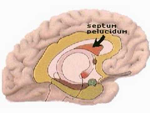
The limbic system is a composite structure which lies in the temporal lobes of the brain and in the region of the thalamus. It is concerned with emotions and memory. The amygdaloid bodies are believed to be concerned with aggression. The hippocampus, lying above the parahippocampal gyrus, is concerned with memory. The septum pelucidum is thought to be associated with pleasure reactions. The cingulate gyrus, the fornix and the anterior commissure carry nerve fibers to and from other structures in the limbic system, as do the mammillary bodies, which are also vitally concerned with memory. These eight areas make up the complete limbic system.

Functions of the Hypothalamus animation

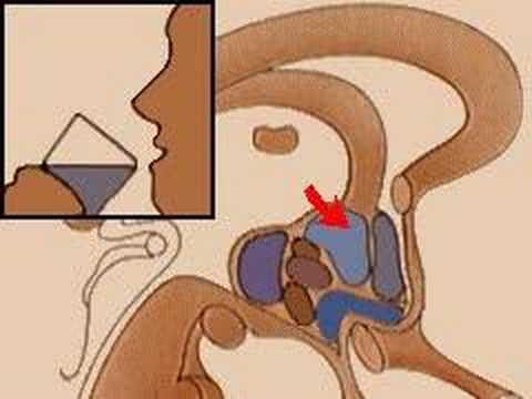
The hypothalamus consists of a number of areas that control the basic drives-hunger, thirst and sex-as well as the internal environment of the body. It is also concerned with emotions and the sensations of pleasure and, possibly, with the sensations of pain and "displeasure". The posterior area controls sexual drives and, therefore, the ability to reproduce the species. The anterior areas control thirst and the drive to find water. The supraoptic nuclei are also concerned with the thirst drive. The preoptic nucleus is the body's thermostat and functions to control internal body temperature. The ventromedial nucleus, or "appestat," controls the hunger drive. The dorsomedial nucleus controls aggressive behavior. The dorsal area is thought to be the human "pleasure center."

Anatomy of the Hypothalamus

Hypothalamus is a portion of the brain that contains a number of small nuclei with a variety of functions. One of the most important functions of the hypothalamus is to link the nervous system to the endocrine system via the pituitary gland (hypophysis). The hypothalamus,  is located below the thalamus, just above the brain stem. In the terminology of neuroanatomy, it forms the ventral part of the diencephalon. All vertebrate brains contain a hypothalamus. In humans, it is roughly the size of an almond.


The hypothalamus is responsible for certain metabolic processes and other activities of the Autonomic Nervous System. It synthesizes and secretes neurohormones, often called hypothalamic-releasing hormones, and these in turn stimulate or inhibit the secretion of pituitary hormones. The hypothalamus controls body temperature, hunger, thirst, fatigue, and circadian cycles.

Cerebellum

The cerebellum (Latin for little brain) is a region of the brain that plays an important role in the integration of sensory perception, coordination and motor control. In order to coordinate motor control, there are many neural pathways linking the cerebellum with the cerebral motor cortex (which sends information to the muscles causing them to move) and the spinocerebellar tract (which provides proprioceptive feedback on the position of the body in space). The cerebellum integrates these pathways, like a train conductor, using the constant feedback on body owning to fine-tune motor movements.


Because of this 'updating' function of the cerebellum, lesions within it are not so debilitating as to cause paralysis, but rather present as feedback deficits resulting in disorders in fine movement, equilibrium, posture, and motor learning. Initial observations by physiologists during the 18th century indicated that patients with cerebellar damage show problems with motor coordination and movement. Research into cerebellar function during the early to mid 19th century was done via lesion and ablation studies in animals. Research physiologists noted that such lesions led to animals with strange movements, awkward gait, and muscular weakness. These observations and studies led to the conclusion that the cerebellum was a motor control structure. However, modern research shows that the cerebellum has a broader role in a number of key cognitive functions, including attention and the processing of language, music, and other sensory temporal stimuli.