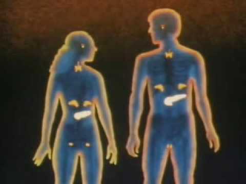
Adrenal glands (also known as suprarenal glands) are the triangle-shaped endocrine glands that sit on top of the kidneys; their name indicates that position (ad-, "near" or "at" + -renes, "kidneys"). They are chiefly responsible for regulating the stress response through the synthesis of corticosteroids and catecholamines, including cortisol and adrenaline.
Anatomically, the adrenal glands are located in the thoracic abdomen situated atop the kidneys, specifically on their anterosuperior aspect. They are also surrounded by the adipose capsule and the renal fascia. In humans, the adrenal glands are found at the level of the 12th thoracic vertebra and receive their blood supply from the adrenal arteries.
The adrenal gland is separated into two distinct structures, both of which receive regulatory input from the nervous system:
Adrenal medulla
As its name suggests, the adrenal medulla is the central core of the adrenal gland, surrounded by the adrenal cortex. The chromaffin cells of the medulla are the body's main source of the catecholamine hormones adrenaline (epinephrine) and noradrenaline (norepinephrine). These water-soluble hormones, derived from the amino acid tyrosine, are part of the fight-or-flight response initiated by the sympathetic nervous system. The adrenal medulla can be considered specialized ganglia of the sympathetic nervous system, lacking distinct synapses, instead releasing secretions directly into the blood. It is also the main source of dopamine, a catecholamine closely related to adrenaline and noradrenaline.
Adrenal cortex
By contrast, the adrenal cortex is devoted to the synthesis of corticosteroid hormones from cholesterol. Some cells belong to the hypothalamic-pituitary-adrenal axis and are the source of cortisol synthesis. Other cortical cells produce androgens such as testosterone, while some regulate water and electrolyte concentrations by secreting aldosterone. In contrast to the direct innervation of the medulla, the cortex is regulated by neuroendocrine hormones secreted by the pituitary gland and hypothalamus, as well as by the renin-angiotensin system.
Arteries and veins
Although variations of the blood supply to the adrenal glands (and indeed the kidneys themselves) are common, there are usually three arteries that supply each adrenal gland:
The superior suprarenal artery is provided by the inferior phrenic
The middle suprarenal artery is provided by the abdominal aorta
The inferior suprarenal artery is provided by the renal artery
Venous drainage of the adrenal glands is achieved via the suprarenal veins:
The right suprarenal vein drains into the inferior vena cava
The left suprarenal vein drains into the left renal vein or the left inferior phrenic vein.
The suprarenal veins receive blood may form anastomoses with the inferior phrenic veins.
The adrenal glands and the thyroid gland are the organs that have the greatest blood supply per gram of tissue. Up to 60 arterioles may enter each adrenal gland.
Hormones are chemicals released by cells that affect cells in other parts of the body. Only a small amount of hormone is required to alter cell metabolism. It is essentially a chemical messenger that transports a signal from one cell to another. All multicellular organisms produce hormones; plant hormones are also called phytohormones. Hormones in animals are often transported in the blood. Cells respond to a hormone when they express a specific receptor for that hormone. The hormone binds to the receptor protein, resulting in the activation of a signal transduction mechanism that ultimately leads to cell type-specific responses.
Endocrine hormone molecules are secreted (released) directly into the bloodstream, while exocrine hormones (or ectohormones) are secreted directly into a duct, and from the duct they either flow into the bloodstream or they flow from cell to cell by diffusion in a process known as paracrine signalling.
Nature of hormonal control
Hormonal regulation of some physiological activities involves a hierarchy of cell types acting on each other either to stimulate or to modulate the release and action of a particular hormone. The secretion of hormones from successive levels of endocrine cells is stimulated by chemical signals originating from cells higher up the hierarchical system. The master coordinator of hormonal activity in mammals is the hypothalamus, which acts on input that it receives from the central nervous system.
Other hormone secretion occurs in response to local conditions, such as the rate of secretion of parathyroid hormone by the parathyroid cells in response to fluctuations of ionized calcium levels in extracellular fluid.
Hormone signaling Hormonal signaling across this hierarchy involves the following: 1. Biosynthesis of a particular hormone in a particular tissue 2. Storage and secretion of the hormone 3. Transport of the hormone to the target cell(s) 4. Recognition of the hormone by an associated cell membrane or intracellular receptor protein. 5. Relay and amplification of the received hormonal signal via a signal transduction process: This then leads to a cellular response. The reaction of the target cells may then be recognized by the original hormone-producing cells, leading to a down-regulation in hormone production. This is an example of a homeostatic negative feedback loop. 6. Degradation of the hormone.
As can be inferred from the hierarchical diagram, hormone biosynthetic cells are typically of a specialized cell type, residing within a particular endocrine gland (e.g., the thyroid gland, the ovaries, or the testes). Hormones may exit their cell of origin via exocytosis or another means of membrane transport. However, the hierarchical model is an oversimplification of the hormonal signaling process. Cellular recipients of a particular hormonal signal may be one of several cell types that reside within a number of different tissues, as is the case for insulin, which triggers a diverse range of systemic physiological effects. Different tissue types may also respond differently to the same hormonal signal. Because of this, hormonal signaling is elaborate and hard to dissect.
Interactions with receptors
Most hormones initiate a cellular response by initially combining with either a specific intracellular or cell membrane associated receptor protein. A cell may have several different receptors that recognize the same hormone and activate different signal transduction pathways, or alternatively different hormones and their receptors may invoke the same biochemical pathway.
For many hormones, including most protein hormones, the receptor is membrane associated and embedded in the plasma membrane at the surface of the cell. The interaction of hormone and receptor typically triggers a cascade of secondary effects within the cytoplasm of the cell, often involving phosphorylation or dephosphorylation of various other cytoplasmic proteins, changes in ion channel permeability, or increased concentrations of intracellular molecules that may act as secondary messengers (e.g. cyclic AMP). Some protein hormones also interact with intracellular receptors located in the cytoplasm or nucleus by an intracrine mechanism.
For hormones such as steroid or thyroid hormones, their receptors are located intracellularly within the cytoplasm of their target cell. In order to bind their receptors these hormones must cross the cell membrane. The combined hormone-receptor complex then moves across the nuclear membrane into the nucleus of the cell, where it binds to specific DNA sequences, effectively amplifying or suppressing the action of certain genes, and affecting protein synthesis. However, it has been shown that not all steroid receptors are located intracellularly, some are plasma membrane associated.
An important consideration, dictating the level at which cellular signal transduction pathways are activated in response to a hormonal signal is the effective concentration of hormone-receptor complexes that are formed. Hormone-receptor complex concentrations are effectively determined by three factors:
1. The number of hormone molecules available for complex formation 2. The number of receptor molecules available for complex formation and 3. The binding affinity between hormone and receptor.
The number of hormone molecules available for complex formation is usually the key factor in determining the level at which signal transduction pathways are activated. The number of hormone molecules available being determined by the concentration of circulating hormone, which is in turn influenced by the level and rate at which they are secreted by biosynthetic cells. The number of receptors at the cell surface of the receiving cell can also be varied as can the affinity between the hormone and its receptor.
Physiology of hormones
Most cells are capable of producing one or more molecules, which act as signaling molecules to other cells, altering their growth, function, or metabolism. The classical hormones produced by cells in the endocrine glands mentioned so far in this article are cellular products, specialized to serve as regulators at the overall organism level. However they may also exert their effects solely within the tissue in which they are produced and originally released.
The rate of hormone biosynthesis and secretion is often regulated by a homeostatic negative feedback control mechanism. Such a mechanism depends on factors which influence the metabolism and excretion of hormones. Thus, higher hormone concentration alone can not trigger the negative feedback mechanism. Negative feedback must be triggered by overproduction of an "effect" of the hormone.
Hormone secretion can be stimulated and inhibited by:
* Other hormones (stimulating- or releasing-hormones) * Plasma concentrations of ions or nutrients, as well as binding globulins * Neurons and mental activity * Environmental changes, e.g., of light or temperature
One special group of hormones is the tropic hormones that stimulate the hormone production of other endocrine glands. For example, thyroid-stimulating hormone (TSH) causes growth and increased activity of another endocrine gland, the thyroid, which increases output of thyroid hormones.
A recently-identified class of hormones is that of the "hunger hormones" - ghrelin, orexin and PYY 3-36 - and "satiety hormones" - e.g., leptin, obestatin, nesfatin-1.
In order to release active hormones quickly into the circulation, hormone biosynthetic cells may produce and store biologically inactive hormones in the form of pre- or prohormones. These can then be quickly converted into their active hormone form in response to a particular stimulus.
The endocrine system is an information signal system much like the nervous system. However, the nervous system uses nerves to conduct information, whereas the endocrine system mainly uses blood vessels as information channels. Glands located in many regions of the body release into the bloodstream specific chemical messengers called hormones. Hormones regulate the many and varied functions of an organism, e.g., mood, growth and development, tissue function, and metabolism, as well as sending messages and acting on them.
The typical mode of cell signaling in the endocrine system is endocrine signaling. However, there are also other modes, i.e., paracrine, autocrine, and neuroendocrine signaling. Purely neurocrine signaling between neurons, on the other hand, belongs completely to the nervous system.
Endocrine
A number of glands that signal each other in sequence is usually referred to as an axis, for example, the hypothalamic-pituitary-adrenal axis.
Typical endocrine glands are the pituitary, thyroid, and adrenal glands. Features of endocrine glands are, in general, their ductless nature, their vascularity, and usually the presence of intracellular vacuoles or granules storing their hormones. In contrast, exocrine glands, such as salivary glands, sweat glands, and glands within the gastrointestinal tract, tend to be much less vascular and have ducts or a hollow lumen.
Autocrine signaling
Autocrine signaling is a form of signalling in which a cell secretes a hormone, or chemical messenger (called the autocrine agent) that binds to autocrine receptors on the same cell, leading to changes in the cell. This can be contrasted with paracrine signaling, intracrine signalling, or classical endocrine signaling.
Paracrine
Paracrine signaling is a form of cell signaling in which the target cell is near ("para" = near) the signal-releasing cell.
A distinction is sometimes made between paracrine and autocrine signaling. Both affect neighboring cells, but whereas autocrine signaling occurs among the same cell, paracrine signaling affects other cells. Two neurons would be an example of a paracrine signal.
Juxtacrine
Juxtacrine signaling is a type of intercellular communication which is transmitted via oligosaccharide, lipid or protein components of a cell membrane and may affect either the emitting cell or immediately adjacent cells.
It occurs between adjacent cells that possess broad patches of closely opposed plasma membrane linked by transmembrane channels known as connexons. The gap between the cells can only usually be between 2-4nm.


 Subscribe in a reader