Pages

Showing posts with label zoology animation. Show all posts

Development of Fetus


A fetus (or foetus or fœtus) is a developing mammal or other viviparous vertebrate, after the embryonic stage and before birth. The plural is fetuses, or sometimes feti.
In humans, the fetal stage of prenatal development begins about eight weeks after fertilization, when the major structures and organ systems have formed, until birth.

The fetal stage begins eight weeks after fertilization. Miscarriage is much less likely at the beginning of the fetal stage.The fetus is not as sensitive to damage from environmental exposures as the embryo was, though toxic exposures can often cause physiological abnormalities or minor congenital malformation. Fetal growth can be terminated by various factors, including miscarriage, feticide committed by a third party, or induced abortion.


Development The following timeline describes some of the specific changes in fetal anatomy and physiology by fertilization age (i.e. the time elapsed since fertilization). Obstetricians often use "gestational age" which, by convention, is measured from 2 weeks earlier than fertilization. For purposes of this article, age is measured from fertilization, except as noted.


8 to 15 weeks The fetal stage commences at eight weeks when the fetus is typically about 30 mm (1.2 inches) in length from crown to rump and the head makes up nearly half of the fetus' size.. The fetus cannot feel pain, is not yet sentient, and moves involuntarily as tissues, organs and pathways begin to develop. The movements include motor patterns, and localized movement of the arms and legs, hiccups, stretches and yawns, sideward bendings of the head, and generalized movements that involve the whole body. These movements are involuntary, and the parts of the fetal brain that control movement will not fully form until late in the second trimester, and the first part of the third trimester. At this stage, the heart is beating but not functional.The hands, feet, head, brain, and other organs are present, but not yet functional. The breathing-like movement of the fetus is necessary for stimulation of lung development, rather than for obtaining oxygen. At nine weeks the fetus' involuntary movements include curling toes to move away from an object, and fingers are structurally able to bend. During weeks 9-12, the face is “well-formed,” though the fetal head is only one to three inches long. From weeks 9 to 12, the fetal eyelids close and remain closed for several months, and the appearance of the genitals in males and females becomes more apparent. Tooth buds appear, the limbs are long and thin, and red blood cells are produced in the liver, however the majority of red blood cells will be made later in gestation (at 21 weeks) by bone marrow. A fine hair called lanugo develops on the head. The gastrointestinal tract, still forming, starts to collect sloughed skin and lanugo, as well as hepatic products, forming meconium (stool). Fetal skin is almost transparent. More muscle tissue and bones have developed, and the bones become harder. The first measurable signs of EEG movement occur in the 12th week. By the end of this stage, the fetus has reached about 15 cm (6 inches). 16 to 25 weeks The lanugo covers the entire body. Eyebrows, eyelashes, fingernails, and toenails appear. The fetus has increased muscle development. Alveoli (air sacs) are forming in lungs. The nervous system develops enough to control some body functions. The cochlea are now developed, though the myelin sheaths in the neural portion of the auditory system will continue to develop until 18 months after birth. The respiratory system has developed to the point where gas exchange is possible. The quickening, the first maternally discernable fetal movements, are often felt during this period. A woman pregnant for the first time (i.e. a primiparous woman) typically feels fetal movements at about 18-19 weeks, whereas a woman who has already given birth at least two times (i.e. a multiparous woman) will typically feel movements around 16 weeks.[22] By the end of the fifth month, the fetus is about 20 cm (8 inches). 26 to 38 weeks The amount of body fat rapidly increases. Lungs are not fully mature. Thalamic brain connections, which mediate sensory input, form. Bones are fully developed, but are still soft and pliable. Iron, calcium, and phosphorus become more abundant. Fingernails reach the end of the fingertips. The lanugo begins to disappear, until it is gone except on the upper arms and shoulders. Small breast buds are present on both sexes. Head hair becomes coarse and thicker. Birth is imminent and occurs around the 38th week. The fetus is considered full-term between weeks 35 and 40,[23] which means that the fetus is considered sufficiently developed for life outside the uterus.[24] It may be 48 to 53 cm (19 to 21 inches) in length, when born.
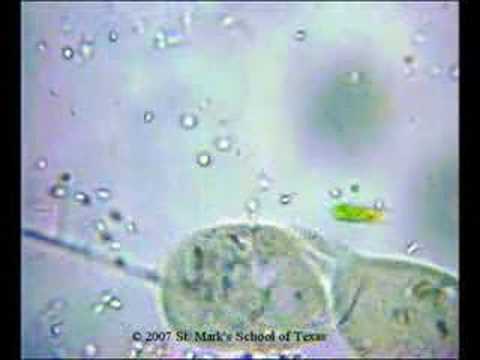
Paramecium Dividing

Paramecia are unicellular organisms usually less than 0.25mm in length and covered with minute hair-like projections called cilia. They are characterized by their cilia which are used in locomotion and during feeding. Paramecia feed on bacteria.


Classification:


Kingdom – Protista
Phylum – Ciliophora
Order – Hymenostomatida
Family -- Paramecidae
Genus – Paramecium
Species – caudatum


Natural Habitat:


Fresh water

Why fresh water?
They take in water by osmosis from the hypotonic environment, bladder-like contractile vacuoles accumulate the excess water from radial canals and periodically expel it through the plasma membrane by contractions of the surrounding cytoplasm.

Interesting characteristics:
Paramecia have 2 nuclei, 1 macronucleus and 1 micronucleus. Some have up to 80 micronuclei!
The organism cannot survive without macronucleus and cannot reproduce without micronucleus.

Reproduction is either by asexual binary fission or occasionally by conjugation (sexual). During binary fission a fully grown organism divides into two daughter cells. Conjugation consists of the temporary union of 2 organisms and the exchange of micronuclear elements. Without the rejuvenating effects of conjugation a paramecium ages and dies. Only opposite mating types, or genetically compatible organisms, can unite in conjugation

Paramecium caudatum in conjugation

Paramecium caudatum are unicellular organisms belonging to the genus of protozoa of the phylum Ciliophora. They are less than 0.25mm in length and covered with minute hair-like projections called cilia. The cilia are used in locomotion and during feeding. They are often called slipper animalcules because of their slipper-like shape.


Paramecium have 2 nuclei (a large macronucleus and a single compact micronucleus). They cannot survive without macro-nucleus and cannot reproduce without micro-nucleus. Reproduction is either by asexual binary fission or occasionally by conjugation (sexual) and rarely by endomixis, a process involving total nuclear reorganization of individual organisms. During binary fission a fully grown organism divides into two daughter cells. Conjugation consists of the temporary union of 2 organisms and the exchange of micro-nuclear elements. Without the rejuvenating effects of conjugation a paramecium ages and dies. Only opposite mating types, or genetically compatible organisms, can unite in conjugation.

Spirotrichs

Spirotrichs are a large and distinctive group of ciliate protozoa. They typically have prominent oral cilia in the form of a series of polykinetids, called the adoral zone of membranelles, beginning anterior to the oral cavity and running down to the left side of the mouth. There may also be one or two paroral membranes on its right side. The body cilia are fused to form polykinetids called cirri in some, and are sparse to absent in others.

Forms with cirri are common throughout soil, freshwater, and marine environments. Individuals tend to be flattened, with cirri confined to the ventral surface. These are variously used for crawling over objects, acting as feet, swimming, or assisting in food capture. They are generally divided into hypotrichs and stichotrichs, but were originally all considered hypotrichs.


Forms with sparse or absent body cilia tend to be smaller and are mostly marine, but a few are common in freshwater. Again, they are generally divided into oligotrichs and choreotrichs, but were originally all considered oligotrichs. The latter group includes the tintinnids, which produce loricae or shells and are the predominant fossil ciliates.

As first defined by Bütschli in 1889 the spirotrichs were one of two orders, together with the now-abandoned holotrichs, and included all ciliates with prominent oral cilia: heterotrichs, hypotrichs, oligotrichs, and peritrichs, although the last were soon separated. The heterotrichs have an adoral zone of membranelles, but molecular and ultrastructure studies have shown they are a separate group that diverged from most other ciliates early on. A few of the smaller groups included with them may be genuine spirotrichs, however, such as the Protocruziida.

The remaining spirotrichs form a monophyletic group, but their relationships are uncertain. For the most part the oligotrichs and choreotrichs appear to form closely related, natural groups. However Halteria and its close relatives, originally considered oligotrichs, form a separate group and may even be modified stichotrichs. Studies also suggest the hypotrichs are paraphyletic to the stichotrichs, and possibly to the oligotrichs and choreotrichs as well. This stands in contrast to the earlier belief that they were the most advanced of all protozoa.

Nerve Physiology - sciatic nerve and gastrocnemius

The sciatic nerve (also known as the ischiatic nerve) is a large nerve that starts in the lower back and runs through the buttock and down the lower limb. It is the longest and widest single nerve in the body.

The sciatic supplies nearly the whole of the skin of the leg, the muscles of the back of the thigh, and those of the leg and foot.The nerve enters the lower limb by exiting the pelvis through the greater sciatic foramen, below the Piriformis muscle and above the superior gemellus muscle.


It descends midway between the greater trochanter of the femur and the tuberosity of the ischium, and along the back of the thigh to about its lower third, where it divides into two large branches, the tibial and common peroneal nerves. This division may take place at any point between the sacral plexus and the lower third of the thigh. When it occurs at the plexus, the common peroneal nerve usually pierces the Piriformis muscles.
Gastrocnemius muscle
Gastrocnemius muscle is a very powerful superficial muscle that is in the back part of the lower leg and also called the calf. It runs from its two heads just above the knee to the heel, and is involved in standing, walking, running and jumping. Along with the soleus muscle it forms the calf muscle.

Hydra feeding

Hydra is a genus of simple, fresh-water animals possessing radial symmetry. Hydras are predatory animals belonging to the phylum Cnidaria and the class Hydrozoa. They can be found in most unpolluted freshwater ponds, lakes and streams in the temperate and tropical regions by gently sweeping a collecting net through weedy areas. They are usually a few milimeters long and are best studied with a microscope. Biologists are especially interested in hydras due to their regenerative ability. Hydras appear to be unique among animals in that they do not undergo senescence (aging).


When feeding, hydras extend their body to maximum length and then slowly extend their tentacles. Despite their simple construction, the tentacles of hydras are extraordinarily extensible and can be four to five times the length of the body. Once fully extended, the tentacles are slowly maneuvered around waiting for contact with a suitable prey animal. Upon contact, nematocysts on the tentacle fire into the prey and the tentacle itself coils around the prey. Within 30 seconds most of the remaining tentacles will have already joined in the attack to subdue the struggling prey. Within two minutes, the tentacles will have surrounded the prey and moved it into the opened mouth aperture. Within ten minutes, the prey will have been enclosed within the gastrovascular cavity and digestion will have started. The hydra is able to stretch its body wall considerably in order to digest prey more than twice its size. After two or three days, the undigestible remains of the prey will be discharged by contractions through the mouth aperture.

The feeding behaviour of the hydra demonstrates the sophistication of what appears to be a simple nervous system.