

Showing posts with label human anatomy. Show all posts
Showing posts with label human anatomy. Show all posts
Systemic and Pulmonary Circulation
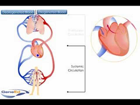
The circulatory system can be viewed as two circuits - the pulmonary circuit and systemic circuit.
Pulmonary circulation involves the transport of blood to and from the lungs. Deoxygenated blood returns to the right side of the heart. This blood is then pumped by the right ventricle into the pulmonary artery (deoxygenated blood) and into the capillaries of the lungs. In the capillaries of the alveoli, oxygen enters the blood and binds to hemoglobin in red blood cells as carbon dioxide diffuses out of the blood into the lungs for removal. The oxygenated blood then travels in the pulmonary vein back to the left atrium of the heart where it joins the systemic circuit.


Systemic circulation involves the transport of blood to and from all the tissues of the body. This circuit is much larger than the pulmonary circuit and so the walls of the left ventricle of the heart are much larger than on the right side. This thicker muscle generates the force required to pump blood all around the systemic circuit. Oxygenated blood is pumped by the left ventricle into the aorta. The aorta then branches into many smaller arteries that carry blood to all areas of the body. In the capillaries, oxygen is delivered to cells and carbon dioxide is picked up for removal. The deoxygenated blood then returns to the anterior vena cava from the upper body and the posterior vena cava from the lower body. Both vessels enter the right atrium of the heart and blood is returned back to the pulmonary circuit for oxygenation.
Function of Cochlea Animation

The Auditory portion of inner ear is called Cochlea,organ of corti is the core component of cochlea which is the sensory organ of hearing, The structure of Cochlea consist of Scala vestibuli (containing perilymp),Scala tympani(containing perilymph),Scala media(containing endolymph),Helicotrema,Reissner's membrane,Basilar membrane ,organ of corti and hair cells,
- Scala vestibulin lies superior to the cochlear duct
- Scala tympani lies inferior to the scala media and terminates at the round window
- Scala media is a membranous duct containing the organ of corti
- Helicotrema is the location where scala tympani and scala vestiduli merge
- Reissner's membrane separates the scala vestibuli from scala media
- Basilar membrane separates the scala media from the scala tympani and determines the mechanical wave propagation properties of the cochlear partion
- Organ of coti is the cellular layer on the basilar membrane,powered by te potential differnce the perilymph and the endolymph
- Hair cells are the sensory cells in the organ of corti
>
When the cochlea uncoils it will be continuous tube divided into 3 separate compartments the Scala media ,Scala tympani ,Scala vestibulin,sound waves are transmitted in scala vestibulin and reach the organ of corti on the Basilar membrane, where stimulate hair like receptor whose tips are fixed in Tectorial membrane


